 Explore
 Validate
 Learn
Learn Immunocytochemistry
 Immunohistochemistry
Antibody data
- Antibody Data
- Antigen structure
- References [2]
- Comments [0]
- Validations
- Immunocytochemistry [2]
- Immunohistochemistry [1]
Submit
Validation data
Reference
Comment
Report error
- Product number
- HPA019589 - Provider product page

- Provider
- Atlas Antibodies
- Proper citation
- Atlas Antibodies Cat#HPA019589, RRID:AB_1853953
- Product name
- Anti-MIER1
- Antibody type
- Polyclonal
- Description
- Polyclonal Antibody against Human MIER1, Gene description: mesoderm induction early response 1, transcriptional regulator, Alternative Gene Names: hMI-ER1, KIAA1610, MI-ER1, Validated applications: IHC, ICC, Uniprot ID: Q8N108, Storage: Store at +4°C for short term storage. Long time storage is recommended at -20°C.
- Reactivity
- Human
- Host
- Rabbit
- Conjugate
- Unconjugated
- Isotype
- IgG
- Vial size
- 100 µl
- Concentration
- 0.1 mg/ml
- Storage
- Store at +4°C for short term storage. Long time storage is recommended at -20°C.
- Handling
- The antibody solution should be gently mixed before use.
Submitted references Acute liver steatosis translationally controls the epigenetic regulator MIER1 to promote liver regeneration in a study with male mice
Role of the BAHD1 Chromatin-Repressive Complex in Placental Development and Regulation of Steroid Metabolism
Chen Y, Chen L, Wu X, Zhao Y, Wang Y, Jiang D, Liu X, Zhou T, Li S, Wei Y, Liu Y, Hu C, Zhou B, Qin J, Ying H, Ding Q
Nature Communications 2023;14(1)
Nature Communications 2023;14(1)
Role of the BAHD1 Chromatin-Repressive Complex in Placental Development and Regulation of Steroid Metabolism
Wade P, Lakisic G, Lebreton A, Pourpre R, Wendling O, Libertini E, Radford E, Le Guillou M, Champy M, Wattenhofer-Donzé M, Soubigou G, Ait-Si-Ali S, Feunteun J, Sorg T, Coppée J, Ferguson-Smith A, Cossart P, Bierne H
PLOS Genetics 2016;12(3):e1005898
PLOS Genetics 2016;12(3):e1005898
No comments: Submit comment
Enhanced validation
Supportive validation
- Submitted by
- 55af80e3e0991
- Enhanced method
- Genetic validation
- Main image
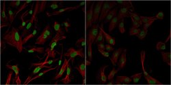
- Experimental details
- Confocal images of immunofluorescently stained human U-2 OS cells.The protein MIER1 is shown in green and the microtubules in red. The image to the left show cells transfected with control siRNA and the image to the right show cells where MIER1 has been downregulated with specific siRNA.
- Sample type
- U-2 OS cells
- Primary Ab dilution
- 1:43
- Secondary Ab
- Secondary Ab
- Secondary Ab dilution
- 1:800
- Knockdown/Genetic Approaches Application
- Immunocytochemistry
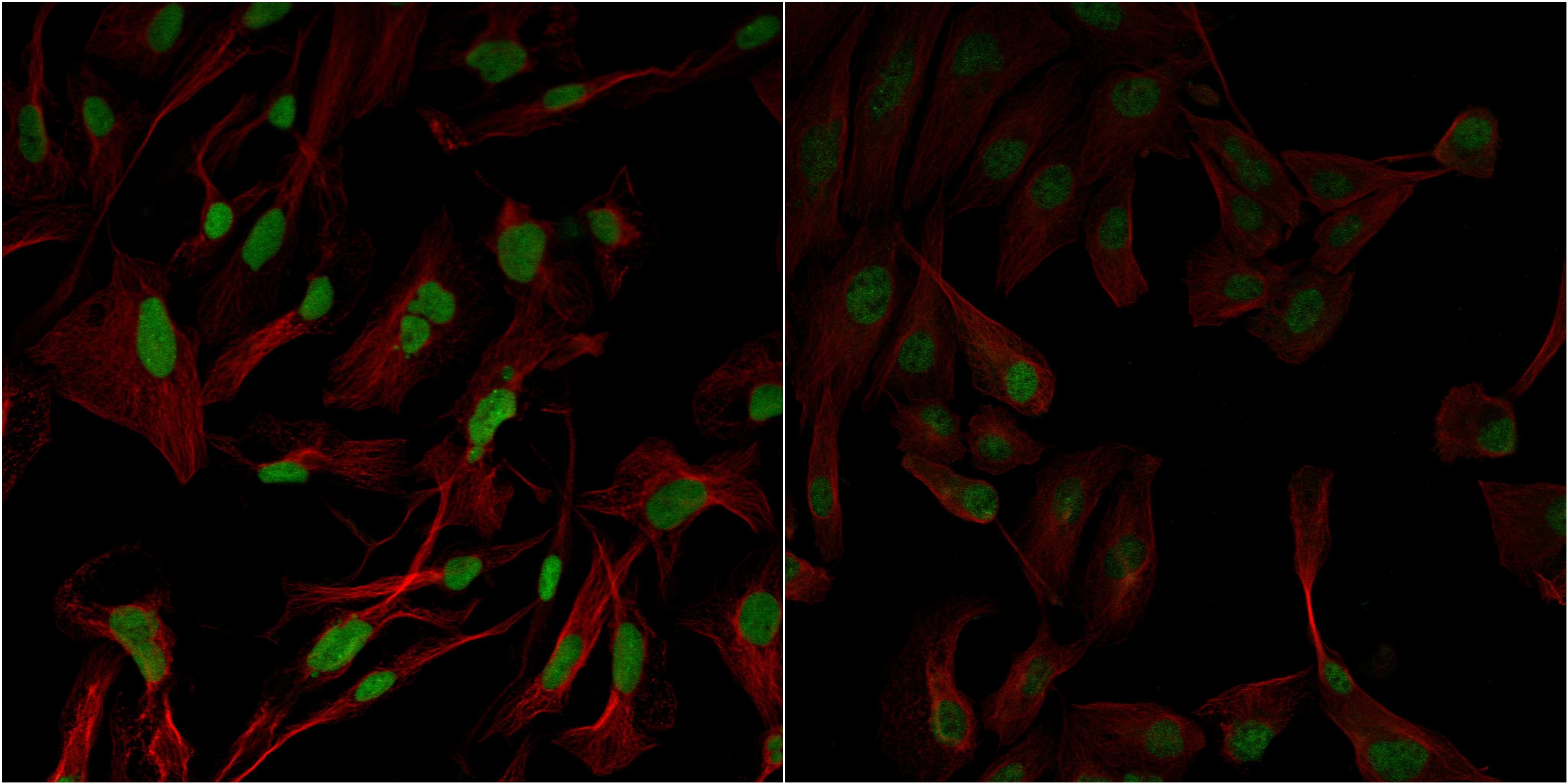
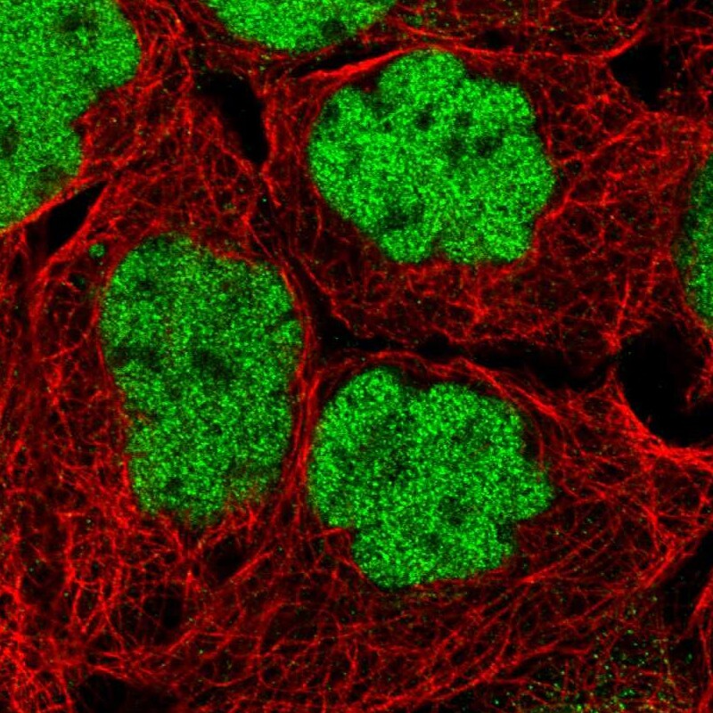
Supportive validation
- Submitted by
- Atlas Antibodies (provider)
- Main image
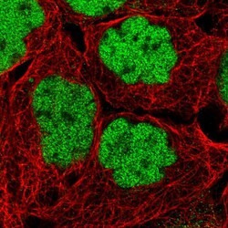
- Experimental details
- Immunofluorescent staining of human cell line A-431 shows localization to nucleoplasm.
- Sample type
- Human
Supportive validation
- Submitted by
- Atlas Antibodies (provider)
- Enhanced method
- Orthogonal validation
- Main image
- Experimental details
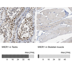
- Immunohistochemistry analysis in human testis and skeletal muscle tissues using HPA019589 antibody. Corresponding MIER1 RNA-seq data are presented for the same tissues.
- Sample type
- Human
- Protocol
- Protocol
