Explore
 Validate
 Learn
Learn Western blot
 Immunocytochemistry
Antibody data
- Antibody Data
- Antigen structure
- References [2]
- Comments [0]
- Validations
- Immunocytochemistry [2]
- Chromatin Immunoprecipitation [2]
- Other assay [1]
Submit
Validation data
Reference
Comment
Report error
- Product number
- PA1-807 - Provider product page

- Provider
- Invitrogen Antibodies
- Product name
- FOXC1 Polyclonal Antibody
- Antibody type
- Polyclonal
- Antigen
- Synthetic peptide
- Description
- PA1-807 detects FOXC1 from human and mouse samples. PA1-807 has been successfully used in Western blot procedures. By Western blot, PA1-807 detects a ~50 kDa band representing FOXC1 from HepG2 cells and mouse kidney cells. This antibody also detects a nonspecific band at ~45 kDa from HepG2 cells, and at ~60 kDa from mouse kidney samples. The PA1-807 immunogen is a synthetic peptide corresponding to residues A(423) V D D P L P D Y S L P(434) of human FOXC1. PA1-807 can be used with blocking peptide PEP-298.
- Reactivity
- Human, Mouse
- Host
- Rabbit
- Isotype
- IgG
- Vial size
- 100 μg
- Concentration
- 1 mg/mL
- Storage
- -20°C, Avoid Freeze/Thaw Cycles
Submitted references Testicular orphan receptor 4 (TR4) promotes papillary thyroid cancer invasion via activating circ-FNLA/miR-149-5p/MMP9 signaling.
System analysis of the regulation of the immune response by CD147 and FOXC1 in cancer cell lines.
Ouyang X, Feng L, Yao L, Xiao Y, Hu X, Zhang G, Liu G, Wang Z
Molecular therapy. Nucleic acids 2021 Jun 4;24:755-767
Molecular therapy. Nucleic acids 2021 Jun 4;24:755-767
System analysis of the regulation of the immune response by CD147 and FOXC1 in cancer cell lines.
Shang YK, Li C, Liu ZK, Kong LM, Wei D, Xu J, Wang ZL, Bian H, Chen ZN
Oncotarget 2018 Feb 27;9(16):12918-12931
Oncotarget 2018 Feb 27;9(16):12918-12931
No comments: Submit comment
Supportive validation
- Submitted by
- Invitrogen Antibodies (provider)
- Main image
- Experimental details
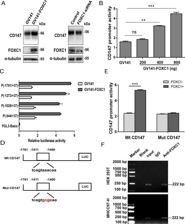
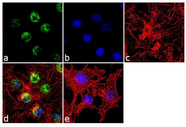
- Immunofluorescence analysis of FOXC1 was done on 70% confluent log phase HepG2 cells. The cells were fixed with 4% paraformaldehyde for 10 minutes, permeabilized with 0.1% Triton™ X-100 for 10 minutes, and blocked with 1% BSA for 1 hour at room temperature. The cells were labeled with FOXC1 Rabbit Polyclonal Antibody (Product # PA1-807) at 2 µg/mL in 0.1% BSA and incubated for 3 hours at room temperature and then labeled with Goat anti-Rabbit IgG (H+L) Superclonal™ Secondary Antibody, Alexa Fluor® 488 conjugate (Product # A27034) at a dilution of 1:2000 for 45 minutes at room temperature (Panel a: green). Nuclei (Panel b: blue) were stained with SlowFade® Gold Antifade Mountant with DAPI (Product # S36938). F-actin (Panel c: red) was stained with Alexa Fluor® 555 Rhodamine Phalloidin (Product # R415, 1:300). Panel d is a merged image showing nuclear localization. Panel e is a no primary antibody control. The images were captured at 60X magnification.
- Submitted by
- Invitrogen Antibodies (provider)
- Main image
- Experimental details
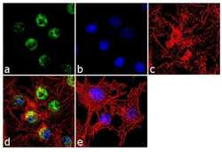
- Immunofluorescence analysis of FOXC1 was done on 70% confluent log phase HepG2 cells. The cells were fixed with 4% paraformaldehyde for 10 minutes, permeabilized with 0.1% Triton™ X-100 for 10 minutes, and blocked with 1% BSA for 1 hour at room temperature. The cells were labeled with FOXC1 Rabbit Polyclonal Antibody (Product # PA1-807) at 2 µg/mL in 0.1% BSA and incubated for 3 hours at room temperature and then labeled with Goat anti-Rabbit IgG (Heavy Chain) Superclonal™ Secondary Antibody, Alexa Fluor® 488 conjugate (Product # A27034) at a dilution of 1:2000 for 45 minutes at room temperature (Panel a: green). Nuclei (Panel b: blue) were stained with SlowFade® Gold Antifade Mountant with DAPI (Product # S36938). F-actin (Panel c: red) was stained with Alexa Fluor® 555 Rhodamine Phalloidin (Product # R415, 1:300). Panel d is a merged image showing nuclear localization. Panel e is a no primary antibody control. The images were captured at 60X magnification.
Supportive validation
- Submitted by
- Invitrogen Antibodies (provider)
- Main image
- Experimental details
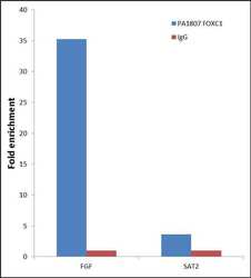
- Enrichment of endogenous FOXC1 Protein at specific gene loci using Anti-FOXC1 Protein Rabbit Polyclonal Antibody: Chromatin Immunoprecipitation (ChIP) was performed using Anti-FOXC1 Protein Rabbit Polyclonal Antibody (Product # PA1-807, 3 µg) on sheared chromatin from 2 million HeLa cells using the "MAGnify ChIP system" kit (Product # 49-2024). Normal Rabbit IgG was used as a negative IP control. The purified DNA was analyzed by 7500 Fast qPCR system (Product # 4351106) with optimized PCR primer pairs for the promoter of active FGF gene, used as positive control target, and the SAT2, used as negative control target. Data is presented as fold enrichment of the antibody signal versus the negative control IgG using the comparative CT method.

- Submitted by
- Invitrogen Antibodies (provider)
- Main image
- Experimental details
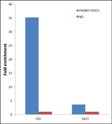
- Enrichment of endogenous FOXC1 Protein at specific gene loci using Anti-FOXC1 Protein Rabbit Polyclonal Antibody: Chromatin Immunoprecipitation (ChIP) was performed using Anti-FOXC1 Protein Rabbit Polyclonal Antibody (Product # PA1-807, 3 µg) on sheared chromatin from 2 million HeLa cells using the "MAGnify ChIP system" kit (Product # 49-2024). Normal Rabbit IgG was used as a negative IP control. The purified DNA was analyzed by 7500 Fast qPCR system (Product # 4351106) with optimized PCR primer pairs for the promoter of active FGF gene, used as positive control target, and the SAT2, used as negative control target. Data is presented as fold enrichment of the antibody signal versus the negative control IgG using the comparative CT method.

Supportive validation
- Submitted by
- Invitrogen Antibodies (provider)
- Main image
- Experimental details
- NULL