 Explore
 Validate
 Learn
Learn Western blot
 Immunocytochemistry
Antibody data
- Antibody Data
- Antigen structure
- References [0]
- Comments [0]
- Validations
- Western blot [1]
- Immunohistochemistry [1]
Submit
Validation data
Reference
Comment
Report error
- Product number
- LS-C348730 - Provider product page

- Provider
- LSBio
- Product name
- Anti-NEUROD1 Antibody (clone BC18-1.11EG) LS-C348730
- Antibody type
- Monoclonal
- Description
- Protein G purified
- Reactivity
- Human
- Host
- Mouse
- Isotype
- IgG
- Antibody clone number
- BC18-1.11EG
- Vial size
- 100µg
- Storage
- Short term 4°C, long term aliquot and store at -20°C, avoid freeze thaw cycles. Store undiluted.
No comments: Submit comment
Supportive validation
- Submitted by
- LSBio (provider)
- Main image
- Experimental details
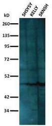
- Figure 3. Shows reactivity of anti-NEUROD1 Antibody (Clone BC18-1.11EG) against SHSY5Y, Kelly and SKNSH neuroblastoma cell lysates by Western blotting. For each cell line, a band of approximately 45kD was observed. This molecular size corresponds to the expected size for NEUROD1. The antibody was used at 2 ug/ml and incubated overnight at 4°C The peroxidase conjugated anti-Mouse Ig secondary antibody was used at 1/10000 dilution and incubated for 2 hours. The blot was developed using enhanced chemiluminescence and image captured on radiographic films.

Supportive validation
- Submitted by
- LSBio (provider)
- Main image
- Experimental details
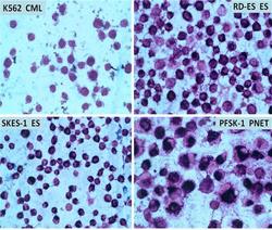
- Figure 2. Shows K562 (chronic myelogenous leukemia), RDES (Ewings sarcoma), SKES (Ewings) sarcoma and PFSK1 (Primitive neuroectodermal tumor) cells stained with anti-NEUROD1 Antibody (Clone BC18-1.11EG) by the indirect immunoperoxidase method. K562 was used as a negative control and showed no staining whereas RDES, SKES-1 and PFSK-1 cells showed weak nuclear but strong cytoplasmic staining. Since the cell lines tested loosely attach to surfaces of chamber slides, the cells were immobilized on adhesion slides for the staining procedure. The cells were fixed in 1% paraformaldehyde, permeabilized in 0.25% Triton X 100 in PBS, blocked with 1% BSA and stained overnight with 5 ug/ml antibody diluted with 1% BSA in PBS. The peroxidase conjugated anti Mouse Ig secondary antibody was used at 1:1000 in 1% BSA in PBS. DAB substrate was used with nickel chloride enhancement to give dark purple/black staining for positive reaction. The substrate was added for 10 minutes, extensively washed, counterstained in diluted hematoxylin, dehydrated, cleared in xylene and mounted.
