 Explore
 Validate
 Learn
LearnPA5-47381
antibody from Invitrogen Antibodies
Targeting: NEUROD1
BETA2, BHF-1, bHLHa3, MODY6, NEUROD
 Western blot
 Immunocytochemistry
Antibody data
- Antibody Data
- Antigen structure
- References [1]
- Comments [0]
- Validations
- Immunocytochemistry [4]
- Chromatin Immunoprecipitation [1]
Submit
Validation data
Reference
Comment
Report error
- Product number
- PA5-47381 - Provider product page

- Provider
- Invitrogen Antibodies
- Product name
- NeuroD1 Polyclonal Antibody
- Antibody type
- Polyclonal
- Antigen
- Recombinant full-length protein
- Description
- Approximately 5% cross-reactivity with recombinant human NeuroD2 is observed. Reconstitute at 0.2 mg/mL in sterile PBS.
- Reactivity
- Human, Mouse
- Host
- Goat
- Isotype
- IgG
- Vial size
- 100 µg
- Concentration
- 0.2 mg/mL
- Storage
- -20° C, Avoid Freeze/Thaw Cycles
Submitted references Abnormal differentiation of stem cells into enteroendocrine cells in rats with DSS-induced colitis.
El-Salhy M, Umezawa K, Hatlebakk JG, Gilja OH
Molecular medicine reports 2017 Apr;15(4):2106-2112
Molecular medicine reports 2017 Apr;15(4):2106-2112
No comments: Submit comment
Supportive validation
- Submitted by
- Invitrogen Antibodies (provider)
- Main image

- Experimental details
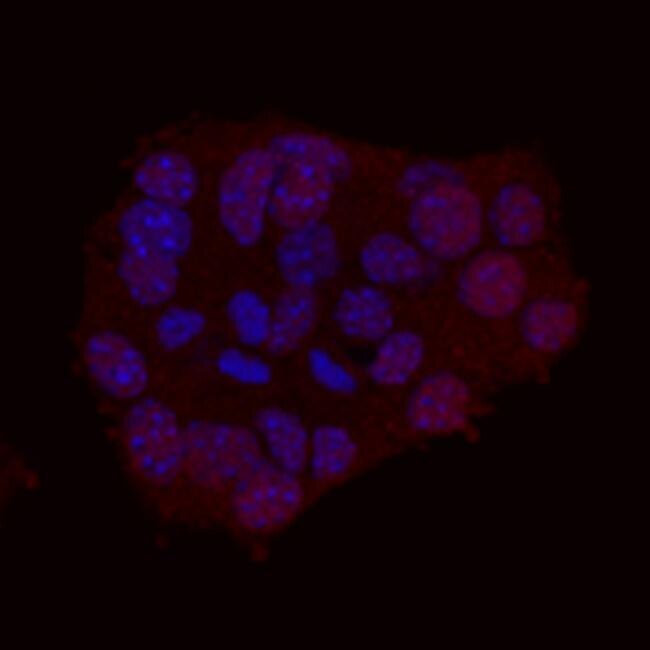
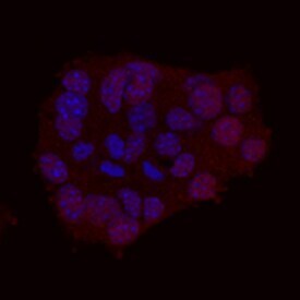
- Immunocytochemical analysis of Neurogenic Differentiation factor 1 (NeuroD1) was detected in immersion fixed ßTC-6 mouse beta cell insulinoma cell line using human/mouse NeuroD1 Antigen Affinity-purified Polyclonal Antibody (Product # PA5-47381) at 10 µg/mL for 3 hours at room temperature. Cells were stained using the 557-conjugated Anti-Goat IgG Secondary Antibody (re and counterstained with DAPI (blue).
- Submitted by
- Invitrogen Antibodies (provider)
- Main image
- Experimental details
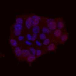
- Immunocytochemistry analysis of NeuroD1 in immersion fixed βTC-6 mouse beta cell insulinoma cell line. Samples were incubated in NeuroD1 polyclonal antibody (Product # PA5-47381) using a dilution of 10 µg/mL for 3 hours at room temperature followed by NorthernLights™ 557-conjugated Anti-Goat IgG Secondary Antibody (red) and counterstained with DAPI (blue).
- Submitted by
- Invitrogen Antibodies (provider)
- Main image

- Experimental details
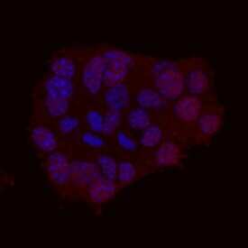
- Immunocytochemistry analysis of NeuroD1 in immersion fixed βTC-6 mouse beta cell insulinoma cell line. Samples were incubated in NeuroD1 polyclonal antibody (Product # PA5-47381) using a dilution of 10 µg/mL for 3 hours at room temperature followed by NorthernLights™ 557-conjugated Anti-Goat IgG Secondary Antibody (red) and counterstained with DAPI (blue).
- Submitted by
- Invitrogen Antibodies (provider)
- Main image
- Experimental details
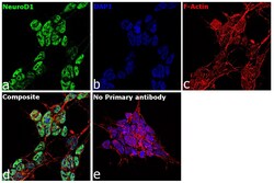
- Immunofluorescence analysis of NeuroD1 was performed using 70% confluent log phase IMR-32 cells. The cells were fixed with 4% paraformaldehyde for 10 minutes, permeabilized with 0.1% Triton™ X-100 for 15 minutes, and blocked with 2% BSA for 1 hour at room temperature. The cells were labeled with NeuroD1 Goat Polyclonal Antibody (Product # PA5-47381) at 5 µg/mL in 0.1% BSA, incubated at 4 degree Celsius overnight and then labeled with Rabbit anti-Goat IgG (H+L) Cross-Adsorbed Secondary Antibody, Alexa Fluor 488 (Product # A-11078) at a dilution of 1:2000 for 45 minutes at room temperature (Panel a: green). Nuclei (Panel b: blue) were stained with ProLong™ Diamond Antifade Mountant with DAPI (Product # P36962). F-actin (Panel c: red) was stained with Rhodamine Phalloidin (Product # R415). Panel d represents the merged image showing Nuclear localization. Panel e represents control cells with no primary antibody to assess background. The images were captured at 60X magnification.
Supportive validation
- Submitted by
- Invitrogen Antibodies (provider)
- Main image
- Experimental details
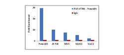
- Chromatin Immunoprecipitation (ChIP) assay of endogenous NeuroD1 protein using Anti-NeuroD1 Antibody: ChIP was performed using Anti-NeuroD1 Polyclonal Antibody (Product # PA5-47381, 2.5 µg) on sheared chromatin from IMR-32 cells using the MAGnify ChIP System kit (Product # 49-2024). Normal Rabbit IgG was used as a negative IP control. The purified DNA was analyzed by qPCR using primers binding to NEUROD1, JUNB, cMYC and SLIT2 transcription start sites and SAT2 satellite repeats. Data is presented as fold enrichment of the antibody signal versus the negative control IgG using the comparative CT method.
