Explore
 Validate
 Learn
LearnPA5-78075
antibody from Invitrogen Antibodies
Targeting: NEUROD1
BETA2, BHF-1, bHLHa3, MODY6, NEUROD
 Western blot
Antibody data
- Antibody Data
- Antigen structure
- References [0]
- Comments [0]
- Validations
- Western blot [4]
- Immunocytochemistry [1]
- Immunohistochemistry [1]
- Chromatin Immunoprecipitation [1]
Submit
Validation data
Reference
Comment
Report error
- Product number
- PA5-78075 - Provider product page

- Provider
- Invitrogen Antibodies
- Product name
- NeuroD1 Polyclonal Antibody
- Antibody type
- Polyclonal
- Antigen
- Synthetic peptide
- Description
- Positive Control: IMR32, IMR32 nuclear extract Predicted Reactivity: Mouse (100%), Bovine (100%) Store product as a concentrated solution. Centrifuge briefly prior to opening the vial.
- Reactivity
- Human, Mouse, Rat
- Host
- Rabbit
- Isotype
- IgG
- Vial size
- 100 µL
- Concentration
- 0.47 mg/mL
- Storage
- Store at 4°C short term. For long term storage, store at -20°C, avoiding freeze/thaw cycles.
No comments: Submit comment
Supportive validation
- Submitted by
- Invitrogen Antibodies (provider)
- Main image
- Experimental details
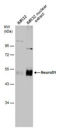
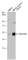
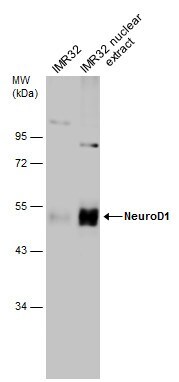
- Western blot analysis of NeuroD1 in whole cell and nuclear lysate using 60 µg of protein. Samples were separated with 7.5% SDS-PAGE and incubated with NeuroD1 polyclonal antibody (Product # PA5-78075) using a dilution of 1:1000.
- Submitted by
- Invitrogen Antibodies (provider)
- Main image
- Experimental details
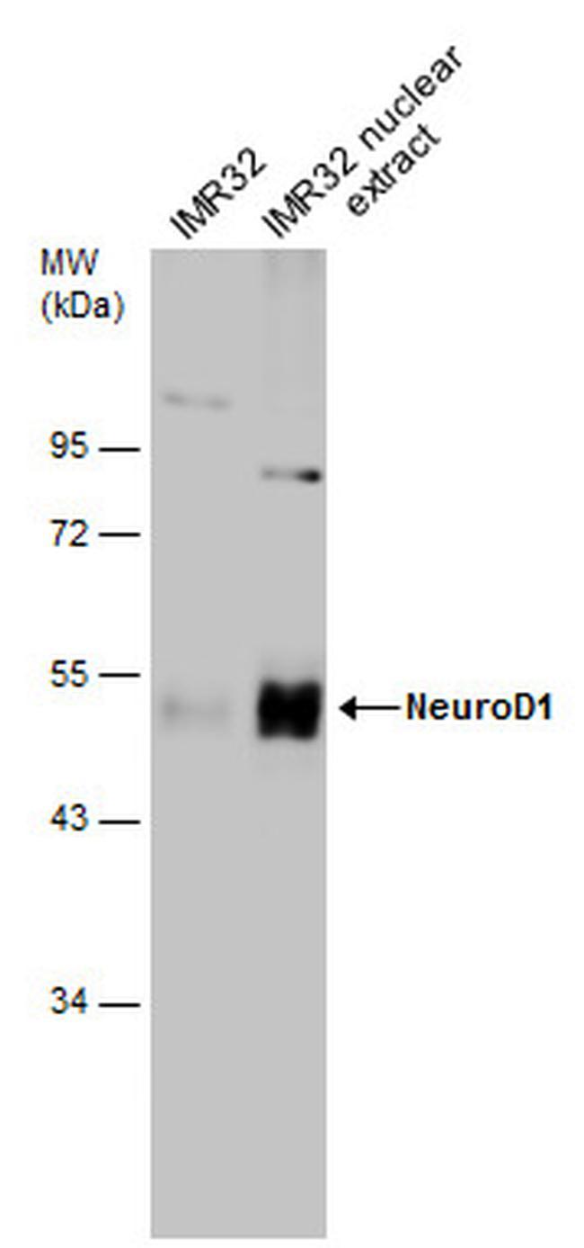
- Western Blot using NeuroD1 Polyclonal Antibody (Product # PA5-78075). IMR32 whole cell and nuclear extracts (60 µg) were separated by 7.5% SDS-PAGE, and the membrane was blotted with NeuroD1 Polyclonal Antibody (Product # PA5-78075) diluted at 1:1,000.
- Submitted by
- Invitrogen Antibodies (provider)
- Main image
- Experimental details
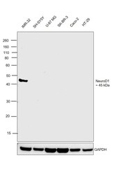
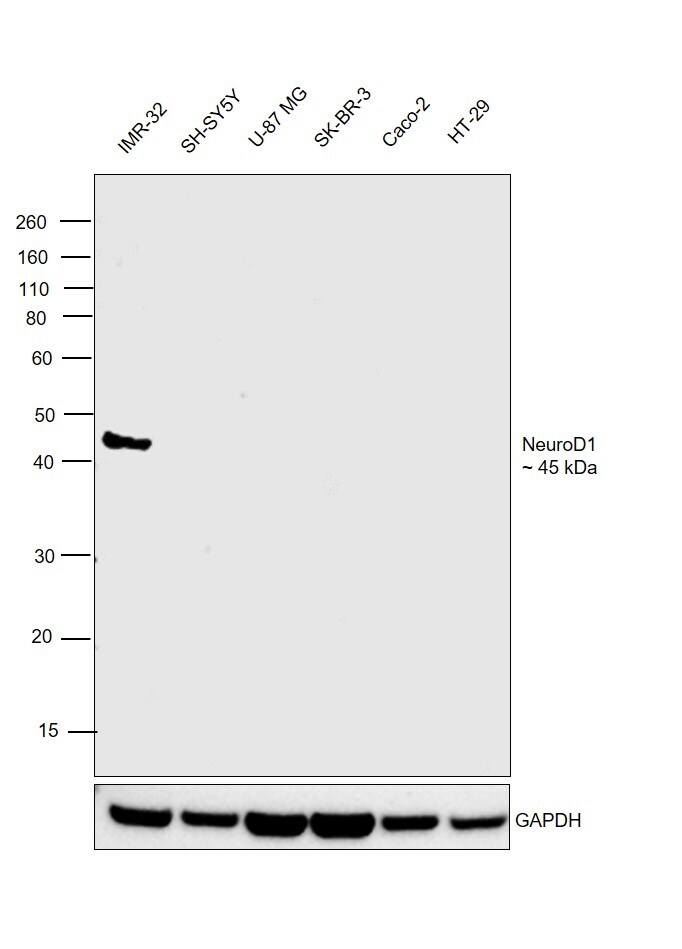
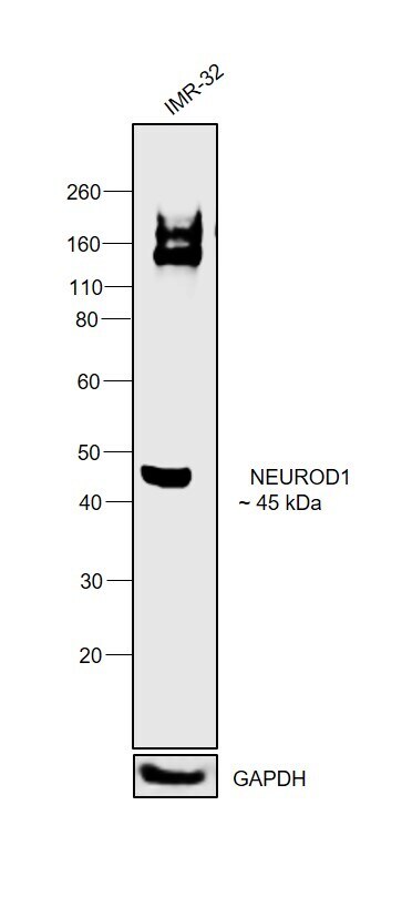
- Western blot was performed using Anti-NeuroD1 Polyclonal Antibody (Product # PA5-78075) and a 45 kDa band corresponding to NEUROD1 was observed only in the IMR-32 cell line, that expresses NeuroD1 at high levels. Nuclear enriched extracts (30 µg lysate) of IMR-32 (Lane 1), SH-SY5Y (Lane 2), U-87 MG (Lane 3), SK-BR-3 (Lane 4), Caco-2 (Lane 5), HT-29 (Lane 6) were electrophoresed using NuPAGE™ 4-12% Bis-Tris Protein Gel (Product # NP0322BOX). Resolved proteins were then transferred onto a nitrocellulose membrane (Product # IB23001) by iBlot® 2 Dry Blotting System (Product # IB21001). The blot was probed with the primary antibody (1:1000 dilution) and detected by chemiluminescence with Goat anti-Rabbit IgG (H+L) Superclonal™ Recombinant Secondary Antibody, HRP (Product # A27036,1:20,000 dilution) using the iBright™ FL1500 Imaging System (Product # A44115). Chemiluminescent detection was performed using SuperSignal™ West Pico PLUS Chemiluminescent Substrate (Product # 34580).
- Submitted by
- Invitrogen Antibodies (provider)
- Main image
- Experimental details
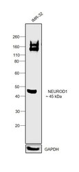
- Western blot was performed using Anti-NeuroD1 Polyclonal Antibody (Product # PA5-78075) and a 45 kDa band corresponding to NEUROD1 was observed in cell line tested. Due to glycosylation a band around 150-230 kDa has also been observed. Modified whole cell extracts (1% SDS) (30 µg lysate) of IMR-32 (Lane 1) were electrophoresed using Novex® NuPAGE® 4-12 % Bis-Tris gel (Product # NP0322BOX). Resolved proteins were then transferred onto a nitrocellulose membrane (Product # IB23001) by iBlot® 2 Dry Blotting System (Product # IB21001). The blot was probed with the primary antibody (1:3000 dilution) and detected by chemiluminescence with Goat anti-Rabbit IgG (H+L), Superclonal™ Recombinant Secondary Antibody, HRP (Product # A27036, 1:4000 dilution) using the iBright FL 1000 (Product # A32752). Chemiluminescent detection was performed using SuperSignal™ West Dura Extended Duration Substrate (Product # 34076).
Supportive validation
- Submitted by
- Invitrogen Antibodies (provider)
- Main image
- Experimental details
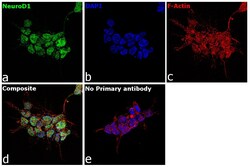
- Immunofluorescence analysis of NeuroD1 was performed using 70% confluent log phase IMR-32. The cells were fixed with 4% paraformaldehyde for 10 minutes, permeabilized with 0.1% Triton™ X-100 for 15 minutes, and blocked with 2% BSA for 1 hour at room temperature. The cells were labeled with NeuroD1 Rabbit Polyclonal Antibody (Product # PA5-78075) at 5 µg/mL in 0.1% BSA, incubated at 4 degree Celsius overnight and then labeled with Goat anti-Rabbit IgG (H+L), Superclonal™ Recombinant Secondary Antibody, Alexa Fluor 488 (Product # A27034) at a dilution of 1:2000 for 45 minutes at room temperature (Panel a: green). Nuclei (Panel b: blue) were stained with ProLong™ Diamond Antifade Mountant with DAPI (Product # P36962). F-actin (Panel c: red) was stained with Rhodamine Phalloidin (Product # R415). Panel d represents the merged image showing Nuclear localization. Panel e represents control cells with no primary antibody to assess background. The images were captured at 60X magnification.

Supportive validation
- Submitted by
- Invitrogen Antibodies (provider)
- Main image

- Experimental details
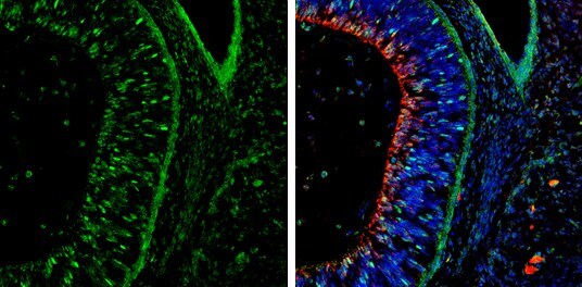
- Immunohistochemistry (Frozen) analysis of NeuroD1 was performed in frozen-sectioned rat E13.5 brain tissue using NeuroD1 Polyclonal Antibody (Product # PA5-78075) at a dilution of 1:250 (Green). Red: beta Tubulin 3/ Tuj1, stained by beta Tubulin 3/ Tuj1 antibody diluted at 1:500. Blue: Fluoroshield with DAPI.
Supportive validation
- Submitted by
- Invitrogen Antibodies (provider)
- Main image

- Experimental details
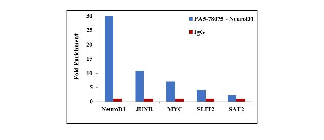
- Chromatin Immunoprecipitation (ChIP) assay of endogenous NeuroD1 protein using Anti-NeuroD1 Antibody: ChIP was performed using Anti-NeuroD1 Rabbit Polyclonal Antibody (Product # PA5-78075, 5 µg) on sheared chromatin from IMR32 cells using the MAGnify ChIP System kit (Product # 49-2024). Normal Rabbit IgG was used as a negative IP control. The purified DNA was analyzed by qPCR using primers binding to NEUROD1, JUNB, cMYC and SLIT2 transcription start sites and SAT2 satellite repeats. Data is presented as fold enrichment of the antibody signal versus the negative control IgG using the comparative CT method.