 Explore
 Validate
 Learn
Learn Western blot
Antibody data
- Antibody Data
- Antigen structure
- References [10]
- Comments [0]
- Validations
- Western blot [2]
- Immunocytochemistry [1]
- Chromatin Immunoprecipitation [1]
Submit
Validation data
Reference
Comment
Report error
- Product number
- AF3797 - Provider product page

- Provider
- R&D Systems
- Product name
- Human/Mouse Smad2/3 Antibody
- Antibody type
- Polyclonal
- Description
- Antigen Affinity-purified. Detects human, mouse, and rat Smad2/3 in Western blots. Predicted to detect rat based on sequence homology.
- Reactivity
- Human, Mouse
- Host
- Goat
- Conjugate
- Unconjugated
- Antigen sequence
P84022- Isotype
- IgG
- Vial size
- 100 ug
- Concentration
- LYOPH
- Storage
- Use a manual defrost freezer and avoid repeated freeze-thaw cycles. 12 months from date of receipt, -20 to -70 °C as supplied. 1 month, 2 to 8 °C under sterile conditions after reconstitution. 6 months, -20 to -70 °C under sterile conditions after reconstitution.
Submitted references COA-Cl prevented TGF-β1-induced CTGF expression by Akt dephosphorylation in normal human dermal fibroblasts, and it attenuated skin fibrosis in mice models of systemic sclerosis.
The SMAD2/3 interactome reveals that TGFβ controls m(6)A mRNA methylation in pluripotency.
Neuronal Protein 3.1 Deficiency Leads to Reduced Cutaneous Scar Collagen Deposition and Tensile Strength due to Impaired Transforming Growth Factor-β1 to -β3 Translation.
Potential mechanisms underlying ectodermal differentiation of Wharton's jelly mesenchymal stem cells.
HEB associates with PRC2 and SMAD2/3 to regulate developmental fates.
miR-373 is regulated by TGFβ signaling and promotes mesendoderm differentiation in human Embryonic Stem Cells.
Antagonism of Nodal signaling by BMP/Smad5 prevents ectopic primitive streak formation in the mouse amnion.
Satellite cell senescence underlies myopathy in a mouse model of limb-girdle muscular dystrophy 2H.
Chromatin and transcriptional signatures for Nodal signaling during endoderm formation in hESCs.
The transforming growth factor-beta/Smad2,3 signalling axis is impaired in experimental pulmonary hypertension.
Nakai K, Karita S, Igarashi J, Tsukamoto I, Hirano K, Kubota Y
Journal of dermatological science 2019 Apr;94(1):205-212
Journal of dermatological science 2019 Apr;94(1):205-212
The SMAD2/3 interactome reveals that TGFβ controls m(6)A mRNA methylation in pluripotency.
Bertero A, Brown S, Madrigal P, Osnato A, Ortmann D, Yiangou L, Kadiwala J, Hubner NC, de Los Mozos IR, Sadée C, Lenaerts AS, Nakanoh S, Grandy R, Farnell E, Ule J, Stunnenberg HG, Mendjan S, Vallier L
Nature 2018 Mar 8;555(7695):256-259
Nature 2018 Mar 8;555(7695):256-259
Neuronal Protein 3.1 Deficiency Leads to Reduced Cutaneous Scar Collagen Deposition and Tensile Strength due to Impaired Transforming Growth Factor-β1 to -β3 Translation.
Cheng T, Yue M, Aslam MN, Wang X, Shekhawat G, Varani J, Schuger L
The American journal of pathology 2017 Feb;187(2):292-303
The American journal of pathology 2017 Feb;187(2):292-303
Potential mechanisms underlying ectodermal differentiation of Wharton's jelly mesenchymal stem cells.
Jadalannagari S, Berry AM, Hopkins RA, Bhavsar D, Aljitawi OS
Biochemical and biophysical research communications 2016 Sep 16;478(2):831-7
Biochemical and biophysical research communications 2016 Sep 16;478(2):831-7
HEB associates with PRC2 and SMAD2/3 to regulate developmental fates.
Yoon SJ, Foley JW, Baker JC
Nature communications 2015 Mar 16;6:6546
Nature communications 2015 Mar 16;6:6546
miR-373 is regulated by TGFβ signaling and promotes mesendoderm differentiation in human Embryonic Stem Cells.
Rosa A, Papaioannou MD, Krzyspiak JE, Brivanlou AH
Developmental biology 2014 Jul 1;391(1):81-8
Developmental biology 2014 Jul 1;391(1):81-8
Antagonism of Nodal signaling by BMP/Smad5 prevents ectopic primitive streak formation in the mouse amnion.
Pereira PN, Dobreva MP, Maas E, Cornelis FM, Moya IM, Umans L, Verfaillie CM, Camus A, de Sousa Lopes SM, Huylebroeck D, Zwijsen A
Development (Cambridge, England) 2012 Sep;139(18):3343-54
Development (Cambridge, England) 2012 Sep;139(18):3343-54
Satellite cell senescence underlies myopathy in a mouse model of limb-girdle muscular dystrophy 2H.
Kudryashova E, Kramerova I, Spencer MJ
The Journal of clinical investigation 2012 May;122(5):1764-76
The Journal of clinical investigation 2012 May;122(5):1764-76
Chromatin and transcriptional signatures for Nodal signaling during endoderm formation in hESCs.
Kim SW, Yoon SJ, Chuong E, Oyolu C, Wills AE, Gupta R, Baker J
Developmental biology 2011 Sep 15;357(2):492-504
Developmental biology 2011 Sep 15;357(2):492-504
The transforming growth factor-beta/Smad2,3 signalling axis is impaired in experimental pulmonary hypertension.
Zakrzewicz A, Kouri FM, Nejman B, Kwapiszewska G, Hecker M, Sandu R, Dony E, Seeger W, Schermuly RT, Eickelberg O, Morty RE
The European respiratory journal 2007 Jun;29(6):1094-104
The European respiratory journal 2007 Jun;29(6):1094-104
No comments: Submit comment
Supportive validation
- Submitted by
- R&D Systems (provider)
- Main image
- Experimental details
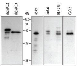
- Detection of Human/Mouse Smad2/3 by Western Blot. Western blot shows lysates of A549 human lung carcinoma cell line, Jurkat human acute T cell leukemia cell line, HEK293 human embryonic kidney cell line, and C2C12 mouse myoblast cell line. PVDF membrane was probed with 0.5 µg/mL Goat Anti-Human/Mouse Smad2/3 Antigen Affinity-purified Polyclonal Antibody (Catalog # AF3797) followed by HRP-conjugated Anti-Goat IgG Secondary Antibody (Catalog # HAF109). For additional reference, recombinant human Smad2 and Smad3 were included. Specific bands for Smad2 were detected at approximately 64 and 58 kDa (as indicated). This experiment was conducted under reducing conditions and using Immunoblot Buffer Group 2.
- Submitted by
- R&D Systems (provider)
- Main image
- Experimental details
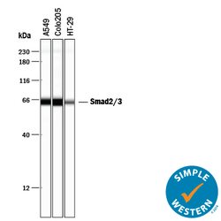
- Detection of Human Smad2/3 by Simple WesternTM. Simple Western lane view shows lysates of A549 human lung carcinoma cell line, COLO 205 human colorectal adenocarcinoma cell line, and HT-29 human colon adenocarcinoma cell line, loaded at 0.2 mg/mL. A specific band was detected for Smad2/3 at approximately 64 kDa (as indicated) using 25 µg/mL of Goat Anti-Human/Mouse Smad2/3 Antigen Affinity-purified Polyclonal Antibody (Catalog # AF3797) followed by 1:50 dilution of HRP-conjugated Anti-Goat IgG Secondary Antibody (Catalog # HAF109). This experiment was conducted under reducing conditions and using the 12-230 kDa separation system.

Supportive validation
- Submitted by
- R&D Systems (provider)
- Main image

- Experimental details
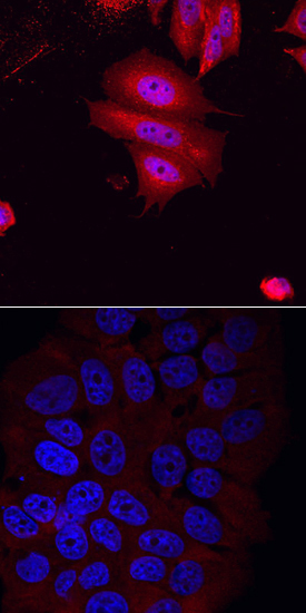
- Smad2/3 in MCF-7 Human Cell Line. Smad2/3 was detected in immersion fixed MCF-7 human breast cancer cell line induced (upper panel) or non-induced (lower panel) to undergo epithelial-mesenchymal transition (EMT) using Goat Anti-Human/Mouse Smad2/3 Antigen Affinity-purified Polyclonal Antibody (Catalog # AF3797) at 10 µg/mL for 3 hours at room temperature. Cells were stained using the NorthernLights™ 557-conjugated Anti-Goat IgG Secondary Antibody (red; Catalog # NL001) and counterstained with DAPI (blue). Specific staining was localized to cytoplasm and, in EMT-induced cells, nuclei. View our protocol for Fluorescent ICC Staining of Cells on Coverslips.
Supportive validation
- Submitted by
- R&D Systems (provider)
- Main image
- Experimental details
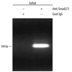
- Detection of Smad2/3-regulated Genes by Chromatin Immunoprecipitation. Jurkat human acute T cell leukemia cell line treated with 50 ng/mL PMA and 200 ng/mL calcium ionomycin for 30 minutes was fixed using formaldehyde, resuspended in lysis buffer, and sonicated to shear chromatin. Smad2/3/DNA complexes were immunoprecipitated using 5 μg Goat Anti-Human/Mouse Smad2/3 Antigen Affinity-purified Polyclonal Antibody (Catalog # AF3797) or control antibody (Catalog # AB-108-C) for 15 minutes in an ultrasonic bath, followed by Biotinylated Anti-Goat IgG Secondary Antibody (Catalog # BAF109). Immunocomplexes were captured using 50 μL of MagCellect Streptavidin Ferrofluid (Catalog # MAG999) and DNA was purified using chelating resin solution. The c-myc promoter was detected by standard PCR.
