 Explore
 Validate
 Learn
Learn Western blot
 ELISA
ELISAAntibody data
- Antibody Data
- Antigen structure
- References [0]
- Comments [0]
- Validations
- Western blot [3]
- ELISA [3]
- Immunohistochemistry [6]
Submit
Validation data
Reference
Comment
Report error
- Product number
- LS-C169142 - Provider product page

- Provider
- LSBio
- Product name
- TBXT / T / Brachyury Antibody (aa 257-309, clone 1H9A2) LS-C169142
- Antibody type
- Monoclonal
- Description
- Purified
- Reactivity
- Human
- Host
- Mouse
- Isotype
- IgG
- Antibody clone number
- 1H9A2
- Storage
- Short term: store at 4°C. Long term: store at -20°C.
No comments: Submit comment
Enhanced validation
- Submitted by
- LSBio (provider)
- Enhanced method
- Genetic validation
- Main image
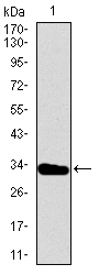
- Experimental details
- Western blot using T monoclonal antibody against human T recombinant protein. (Expected MW is 31.2 kDa)
- Submitted by
- LSBio (provider)
- Enhanced method
- Genetic validation
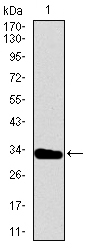
- Main image

- Experimental details
- Western blot using T monoclonal antibody against HEK293 (1) and T (AA: 257-309)-hIgGFc transfected HEK293 (2) cell lysate.
- Submitted by
- LSBio (provider)
- Enhanced method
- Genetic validation
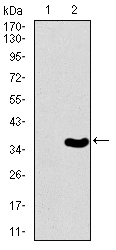
- Main image

- Experimental details
- Western blot using T mouse monoclonal antibody against Raji (1), and Jurkat (2) cell lysate.
Supportive validation
- Submitted by
- LSBio (provider)
- Enhanced method
- Genetic validation
- Main image

- Experimental details
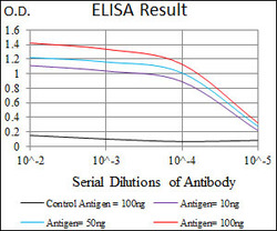
- Red: Control Antigen (100ng); Purple: Antigen (10ng); Green: Antigen (50ng); Blue: Antigen (100ng);

- Submitted by
- LSBio (provider)
- Main image
- Experimental details
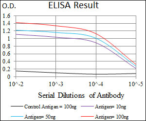
- Red: Control Antigen (100ng); Purple: Antigen (10ng); Green: Antigen (50ng); Blue: Antigen (100ng);
- Submitted by
- LSBio (provider)
- Main image

- Experimental details
- Red: Control Antigen (100ng); Purple: Antigen (10ng); Green: Antigen (50ng); Blue: Antigen (100ng);

Enhanced validation
- Submitted by
- LSBio (provider)
- Enhanced method
- Genetic validation
- Main image

- Experimental details
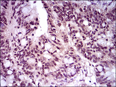
- IHC of paraffin-embedded cervical cancer tissues using T mouse monoclonal antibody with DAB staining.

- Submitted by
- LSBio (provider)
- Enhanced method
- Genetic validation
- Main image

- Experimental details
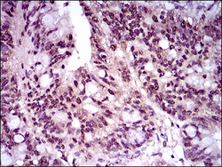
- IHC of paraffin-embedded rectum cancer tissues using T mouse monoclonal antibody with DAB staining.
- Submitted by
- LSBio (provider)
- Enhanced method
- Genetic validation
- Main image

- Experimental details
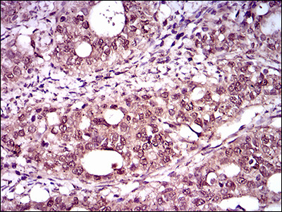
- IHC of paraffin-embedded cervical cancer tissues using T mouse monoclonal antibody with DAB staining.

- Submitted by
- LSBio (provider)
- Enhanced method
- Genetic validation
- Main image
- Experimental details
- IHC of paraffin-embedded rectum cancer tissues using T mouse monoclonal antibody with DAB staining.

- Submitted by
- LSBio (provider)
- Main image

- Experimental details
- IHC of paraffin-embedded cervical cancer tissues using T mouse monoclonal antibody with DAB staining.
- Submitted by
- LSBio (provider)
- Main image

- Experimental details
- IHC of paraffin-embedded rectum cancer tissues using T mouse monoclonal antibody with DAB staining.
