 Explore
 Validate
 Learn
Learn Western blot
Antibody data
- Antibody Data
- Antigen structure
- References [0]
- Comments [0]
- Validations
- Western blot [3]
- ELISA [1]
- Immunohistochemistry [2]
- Flow cytometry [1]
Submit
Validation data
Reference
Comment
Report error
- Product number
- NBP2-37434 - Provider product page

- Provider
- Novus Biologicals
- Product name
- Mouse Monoclonal Brachyury Antibody
- Antibody type
- Monoclonal
- Description
- Protein A or G purified.
- Reactivity
- Human
- Host
- Mouse
- Isotype
- IgG
- Vial size
- 0.1 ml
- Concentration
- 1 mg/ml
- Storage
- Store at 4C short term. Aliquot and store at -20C long term. Avoid freeze-thaw cycles.
No comments: Submit comment
Supportive validation
- Submitted by
- Novus Biologicals (provider)
- Main image
- Experimental details
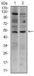
- Western Blot: Brachyury Antibody (1H9A2) [NBP2-37434] - Western blot analysis using T mouse mAb against Raji (1), and Jurkat (2) cell lysate.

- Submitted by
- Novus Biologicals (provider)
- Main image
- Experimental details
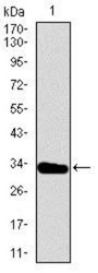
- Western Blot: Brachyury Antibody (1H9A2) [NBP2-37434] - Western blot analysis using T mAb against human T recombinant protein. (Expected MW is 31.2 kDa)

- Submitted by
- Novus Biologicals (provider)
- Main image
- Experimental details
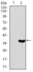
- Western Blot: Brachyury Antibody (1H9A2) [NBP2-37434] - Western blot analysis using T mAb against HEK293 (1) and T (AA: 257-309)-hIgGFc transfected HEK293 (2) cell lysate.

Supportive validation
- Submitted by
- Novus Biologicals (provider)
- Main image

- Experimental details
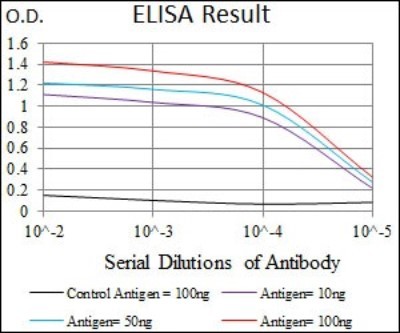
- ELISA: Brachyury Antibody (1H9A2) [NBP2-37434] - Red: Control Antigen (100ng); Purple: Antigen (10ng); Green: Antigen (50ng); Blue: Antigen (100ng);
Supportive validation
- Submitted by
- Novus Biologicals (provider)
- Main image
- Experimental details
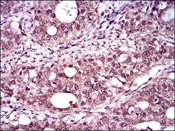
- Immunohistochemistry-Paraffin: Brachyury Antibody (1H9A2) [NBP2-37434] - Analysis of cancer tissues using T mouse mAb with DAB staining.

- Submitted by
- Novus Biologicals (provider)
- Main image

- Experimental details
- Immunohistochemistry: Brachyury Antibody (1H9A2) [NBP2-37434] - Analysis of rectum cancer tissues using T mouse mAb with DAB staining.

Supportive validation
- Submitted by
- Novus Biologicals (provider)
- Main image
- Experimental details
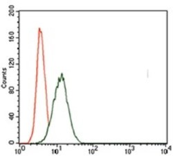
- Flow Cytometry: Brachyury Antibody (1H9A2) [NBP2-37434] - Flow cytometric analysis of HeLa cells using T mouse mAb (green) and negative control (red).
