Explore
 Validate
 Learn
Learn Western blot
 Immunocytochemistry
Antibody data
- Antibody Data
- Antigen structure
- References [0]
- Comments [0]
- Validations
- Immunocytochemistry [5]
- Immunohistochemistry [1]
- Chromatin Immunoprecipitation [2]
Submit
Validation data
Reference
Comment
Report error
- Product number
- PA5-46984 - Provider product page

- Provider
- Invitrogen Antibodies
- Product name
- Brachyury Polyclonal Antibody
- Antibody type
- Polyclonal
- Antigen
- Recombinant full-length protein
- Description
- In direct ELISAs, approximately 25% cross-reactivity with recombinant human (rh) TBX-6 is observed, and approximately 5% cross-reactivity with rhTBX-2, rhTBX-5, and rhTBX-18 is observed. Reconstitute at 0.2 mg/mL in sterile PBS.
- Reactivity
- Human, Mouse
- Host
- Goat
- Isotype
- IgG
- Vial size
- 100 μg
- Concentration
- 0.2 mg/mL
- Storage
- -20°C, Avoid Freeze/Thaw Cycles
No comments: Submit comment
Supportive validation
- Submitted by
- Invitrogen Antibodies (provider)
- Main image
- Experimental details
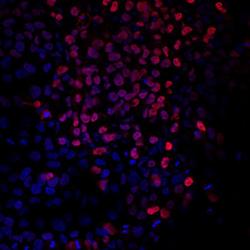
- Immunocytochemical analysis of Brachyury was detected in immersion fixed BG01V human embryonic stem cells differentiated into mesoderm using Goat Anti-human Brachyury Antigen Affinity-purified Polyclonal Antibody (Product # PA5-46984) at 10 µg/mL for 3 hours at room temperature. Cells were stained using a 557-conjugated Anti-Goat IgG Secondary Antibody (re and counterstained with DAPI (blue). Specific staining was localized to nuclei.
- Submitted by
- Invitrogen Antibodies (provider)
- Main image

- Experimental details
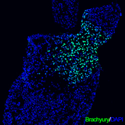
- Immunocytochemistry analysis of Brachyury in immersion fixed differentiated human embryonic stem cells. Samples were incubated in Brachyury polyclonal antibody (Product # PA5-46984) using a dilution of 10 µg/mL for 3 hours at room temperature. Cells were stained (green) and counterstained with DAPI (blue).

- Submitted by
- Invitrogen Antibodies (provider)
- Main image

- Experimental details
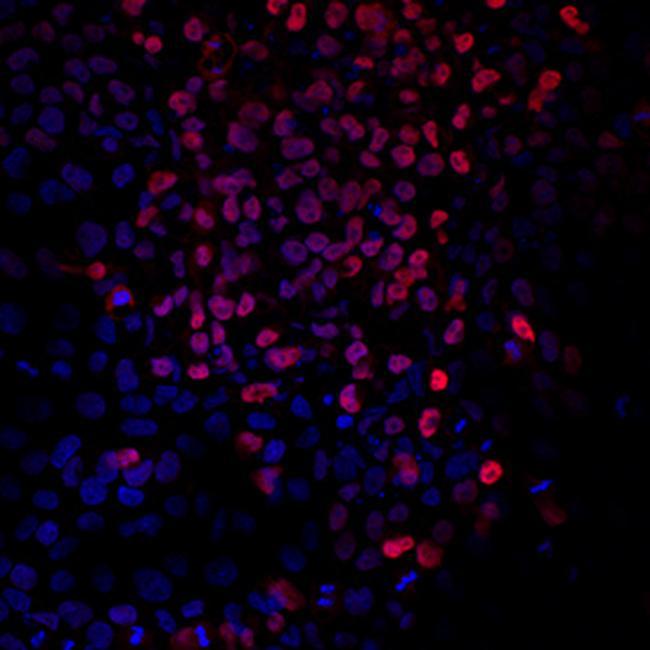
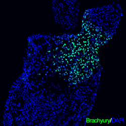
- Immunocytochemistry analysis of Brachyury in immersion fixed BG01V human embryonic stem cells differentiated into mesoderm. Samples were incubated in Brachyury polyclonal antibody (Product # PA5-46984) using a dilution of 10 µg/mL for 3 hours at room temperature followed by NorthernLights™ 557-conjugated Anti-Goat IgG Secondary Antibody (red) and counterstained with DAPI (blue). Specific staining was localized to nuclei.

- Submitted by
- Invitrogen Antibodies (provider)
- Main image
- Experimental details
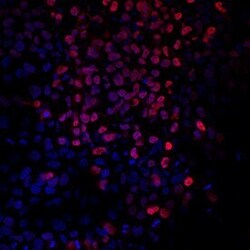
- Immunocytochemistry analysis of Brachyury in immersion fixed differentiated human embryonic stem cells. Samples were incubated in Brachyury polyclonal antibody (Product # PA5-46984) using a dilution of 10 µg/mL for 3 hours at room temperature. Cells were stained (green) and counterstained with DAPI (blue).
Supportive validation
- Submitted by
- Invitrogen Antibodies (provider)
- Main image

- Experimental details
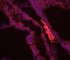
- Immunohistochemical analysis of Brachyury in immersion fixed frozen sections of embryonic mouse notochord (E9.5). Samples were incubated in Brachyury polyclonal antibody (Product # PA5-46984) using a dilution of 10 µg/mL overnight at 4 °C followed by NorthernLights™ 557-conjugated Anti-Goat IgG Secondary Antibody (red) and counterstained with DAPI (blue).
Supportive validation
- Submitted by
- Invitrogen Antibodies (provider)
- Main image
- Experimental details
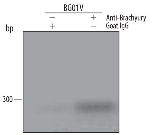
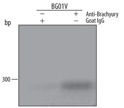
- ChIP assay of Brachyury in Mesoderm-differentiated BG01V human embryonic stem cells. Samples were immunoprecipitated with Brachyury polyclonal antibody (Product # PA5-46984) for 15 minutes in an ultrasonic bath using a dilution of 5 μg followed by a Biotinylated Anti-Goat IgG secondary antibody. Cells were fixed using formaldehyde, resuspended in lysis buffer, and sonicated to shear chromatin. Immunocomplexes were captured using 50 μL of MagCellect Streptavidin Ferrofluid and DNA was purified using chelating resin solution. The VEGF promoter was detected by standard PCR.

- Submitted by
- Invitrogen Antibodies (provider)
- Main image

- Experimental details
- ChIP assay of Brachyury in Mesoderm-differentiated BG01V human embryonic stem cells. Samples were immunoprecipitated with Brachyury polyclonal antibody (Product # PA5-46984) for 15 minutes in an ultrasonic bath using a dilution of 5 μg followed by a Biotinylated Anti-Goat IgG secondary antibody. Cells were fixed using formaldehyde, resuspended in lysis buffer, and sonicated to shear chromatin. Immunocomplexes were captured using 50 μL of MagCellect Streptavidin Ferrofluid and DNA was purified using chelating resin solution. The VEGF promoter was detected by standard PCR.