Explore
 Validate
 Learn
Learn Western blot
 ELISA
ELISAAntibody data
- Antibody Data
- Antigen structure
- References [1]
- Comments [0]
- Validations
- Western blot [3]
Submit
Validation data
Reference
Comment
Report error
- Product number
- GTX24731 - Provider product page

- Provider
- GeneTex
- Proper citation
- GeneTex Cat#GTX24731, RRID:AB_423851
- Product name
- WHIP antibody
- Antibody type
- Polyclonal
- Reactivity
- Human, Mouse, Rat, Simian
- Host
- Rabbit
Submitted references Human Wrnip1 is localized in replication factories in a ubiquitin-binding zinc finger-dependent manner.
Crosetto N, Bienko M, Hibbert RG, Perica T, Ambrogio C, Kensche T, Hofmann K, Sixma TK, Dikic I
The Journal of biological chemistry 2008 Dec 12;283(50):35173-85
The Journal of biological chemistry 2008 Dec 12;283(50):35173-85
No comments: Submit comment
Supportive validation
- Submitted by
- GeneTex (provider)
- Main image
- Experimental details
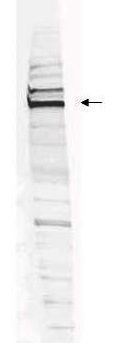
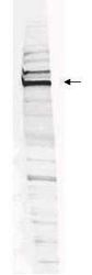
- Western blot analysis is shown using GeneTex Affinity Purified anti-Human WHIP antibody (GTX24731) to detect Human WHIP present in a HEK293 whole cell lysate. ~30mg of lysate was loaded per lane for 4-20% gradient SDS-PAGE. Comparison to a molecular weight marker (not shown) indicates a primary band of ~96.0 kDa is detected. The identity of the minor band migrating at a slightly higher molecular weight is unknown, but may represent an alternate isoform of WHIP or post translational modification of the WHIP protein. See Figure 2 for the results of peptide competition experiments. The blot was incubated with a 1:200 dilution of the antibody at room temperature for 2 h followed by detection using infrared labeled Goat-a-Rabbit IgG [H&L] MX10 diluted 1:5,000 for 45 min. The fluorescence image was captured using the Odyssey® Infrared Imaging System developed by LI-COR.
- Validation comment
- WB
- Submitted by
- GeneTex (provider)
- Main image
- Experimental details
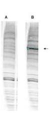
- Western blot analysis is shown using GeneTex anti-Human WHIP antibody (GTX24731) with and without pre-incubation with blocking peptide. Testing was performed on antiserum prior to affinity purification. Peptide competition (left) blocks the specific staining, whereas the control (right) shows staining of a strong dominant band corresponding to human WHIP1. ~30?g of HEK293 lysate was loaded per lane for 4-20% gradient SDS-PAGE. Comparison to a molecular weight marker (not shown) indicates a band of ~96.0 kDa is detected. The blot was incubated with a 1:1000 dilution of the antibody at room temperature for 2 h followed by detection using infrared labeled Goat-a-Rabbit IgG [H&L] MX10 diluted 1:5,000 for 45 min.The fluorescence image was captured using the Odyssey® Infrared Imaging System developed by LI-COR.
- Validation comment
- WB

- Submitted by
- GeneTex (provider)
- Main image
- Experimental details
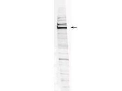
- Western blot analysis is shown using GeneTex Affinity Purified anti-Human WHIP antibody (GTX24731) to detect Human WHIP present in a HEK293 whole cell lysate. ~30mg of lysate was loaded per lane for 4-20% gradient SDS-PAGE. Comparison to a molecular weight marker (not shown) indicates a primary band of ~96.0 kDa is detected. The identity of the minor band migrating at a slightly higher molecular weight is unknown, but may represent an alternate isoform of WHIP or post translational modification of the WHIP protein. The blot was incubated with a 1:200 dilution of the antibody at room temperature for 2 h followed by detection using infrared labeled Goat-a-Rabbit IgG [H&L] MX10 diluted 1:5,000 for 45 min. The fluorescence image was captured using the Odyssey? Infrared Imaging System developed by LI-COR.