 Explore
 Validate
 Learn
LearnAP09250PU-N
antibody from Acris Antibodies GmbH
Targeting: WRNIP1
bA420G6.2, CFAP93, FAP93, FLJ22526, WHIP
 Western blot
 ELISA
ELISAAntibody data
- Antibody Data
- Antigen structure
- References [0]
- Comments [0]
- Validations
- Western blot [2]
Submit
Validation data
Reference
Comment
Report error
- Product number
- AP09250PU-N - Provider product page

- Provider
- Acris Antibodies GmbH
- Proper citation
- Acris Antibodies GmbH Cat#AP09250PU-N, RRID:AB_2035153
- Product name
- anti ATPase WRNIP1
- Antibody type
- Polyclonal
- Antigen
- Synthetic peptide corresponding to an internal region of the WHIP1 protein
- Reactivity
- Human, Mouse, Rat, Simian
- Host
- Rabbit
- Isotype
- IgG
- Vial size
- 0.1 mg
- Concentration
- 1.10 mg/ml (by UV absorbance at 280 nm)
No comments: Submit comment
Supportive validation
- Submitted by
- Acris Antibodies GmbH (provider)
- Main image
- Experimental details
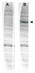
- Western blot analysis is shown using WHIP antibody with and without pre-incubation with blocking peptide. Testing was performed on antiserum prior to affinity purification. Peptide competition (left) blocks the specific staining, whereas the control (right) shows staining of a strong dominant band corresponding to human WHIP1. ~30μg of HEK293 lysate was loaded per lane for 4-20% gradient SDS-PAGE. Comparison to a molecular weight marker (not shown) indicates a band of ~96.0 kDa is detected. The blot was incubated with a 1:1000 dilution of the antibody at room temperature for 2 h followed by detection using IRDye® 800 labeled Goata-Rabbit IgG [H&L] MX10 diluted 1:5,000 for 45 min. IRDye® 800 fluorescence image was captured using the Odyssey® Infrared Imaging System developed by LI-COR. IRDye is a trademark of LI-COR, Inc. Other systems will yield similar results.

- Submitted by
- Acris Antibodies GmbH (provider)
- Main image
- Experimental details
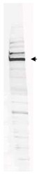
- Western blot analysis is shown using anti-Human WHIP antibody to detect Human WHIP present in a HEK293 whole cell lysate. ~30μg of lysate was loaded per lane for 4-20% gradient SDSPAGE. Comparison to a molecular weight marker (not shown) indicates a primary band of ~96.0 kDa is detected. The identity of the minor band migrating at a slightly higher molecular weight is unknown, but may represent an alternate isoform of WHIP or post translational modification of the WHIP protein. See Figure 2 for the results of peptide competition experiments. The blot was incubated with a 1:200 dilution of the antibody at room temperature for 2 h followed by detection using IRDye® 800 labeled Goat-a-Rabbit IgG [H&L] MX10 (611-132-122) diluted 1:5,000 for 45 min. IRDye® 800 fluorescence image was captured using the Odyssey®Infrared Imaging System developed by LI-COR. IRDye is a trademark of LI-COR, Inc. Other detection systems will yield similar results.
