 Explore
 Validate
 Learn
LearnNBP1-77987
antibody from Novus Biologicals
Targeting: WRNIP1
bA420G6.2, CFAP93, FAP93, FLJ22526, WHIP
 Western blot
 ELISA
ELISAAntibody data
- Antibody Data
- Antigen structure
- References [0]
- Comments [0]
- Validations
- Western blot [2]
Submit
Validation data
Reference
Comment
Report error
- Product number
- NBP1-77987 - Provider product page

- Provider
- Novus Biologicals
- Proper citation
- Novus Cat#NBP1-77987, RRID:AB_11035326
- Product name
- Rabbit Polyclonal WHIP Antibody
- Antibody type
- Polyclonal
- Description
- Immunogen affinity purified. Reactivity is expected against mouse, rat and monkey WHIP1 protein. The immunogen sequence shows 100% homology to human WHIP1 (isoform 1) and WHIP2 (isoform 2) with predicted molecular weights of 72.2 kDa and 69.5 kDa, respectively. Reactivity with WHIP proteins from other sources is not known, but is likely due to reported homologies.
- Reactivity
- Human, Mouse, Rat
- Host
- Rabbit
- Isotype
- IgG
- Vial size
- 0.1 mg
- Concentration
- 1 mg/ml
- Storage
- Store at -20C. Avoid freeze-thaw cycles.
No comments: Submit comment
Supportive validation
- Submitted by
- Novus Biologicals (provider)
- Main image
- Experimental details
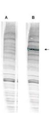
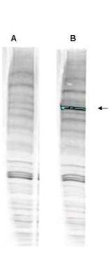
- Western Blot: WHIP Antibody [NBP1-77987] - Antibody with and without pre-incubation with blocking peptide. Testing was performed on antiserum prior to affinity purification. Peptide competition (left) blocks the specific staining, whereas the control (right) shows staining of a strong dominant band corresponding to human WHIP1. ~30ug of HEK293 lysate was loaded per lane for 4-20% gradient SDS-PAGE. Comparison to a molecular weight marker (not shown) indicates a band of ~96.0 kDa is detected. The blot was incubated with a 1:1000 dilution of the antibody at room temperature for 2 h followed by detection using IRDye(R) 800 labeled Goat-a-Rabbit IgG [H&L] MX10 diluted 1:5,000 for 45 min.
- Submitted by
- Novus Biologicals (provider)
- Main image
- Experimental details
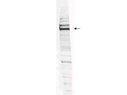
- Western Blot: WHIP Antibody [NBP1-77987] - HEK293 whole cell lysate. ~30ug of lysate was loaded per lane for 4-20% gradient SDS-PAGE. Comparison to a molecular weight marker (not shown) indicates a primary band of ~96.0 kDa is detected. The identity of the minor band migrating at a slightly higher molecular weight is unknown, but may represent an alternate isoform of WHIP or post translational modification of the WHIP protein. See Figure 2 for the results of peptide competition experiments. The blot was incubated with a 1:200 dilution of the antibody at room temperature for 2 h followed by detection using IRDye(R) 800 labeled Goat-a-Rabbit IgG [H&L] MX10 diluted 1:5,000 for 45 min.
