Explore
 Validate
 Learn
Learn Western blot
Antibody data
- Antibody Data
- Antigen structure
- References [2]
- Comments [0]
- Validations
- Western blot [1]
- Immunohistochemistry [1]
Submit
Validation data
Reference
Comment
Report error
- Product number
- AF5045 - Provider product page

- Provider
- R&D Systems
- Product name
- Human/Mouse MSX1 Antibody
- Antibody type
- Polyclonal
- Description
- Antigen Affinity-purified. Detects human and mouse MSX1 in Western blots.
- Reactivity
- Human, Mouse
- Host
- Goat
- Conjugate
- Unconjugated
- Antigen sequence
P28360- Isotype
- IgG
- Vial size
- 100 ug
- Concentration
- LYOPH
- Storage
- Use a manual defrost freezer and avoid repeated freeze-thaw cycles. 12 months from date of receipt, -20 to -70 °C as supplied. 1 month, 2 to 8 °C under sterile conditions after reconstitution. 6 months, -20 to -70 °C under sterile conditions after reconstitution.
Submitted references Msx1 loss suppresses formation of the ectopic crypts developed in the Apc-deficient small intestinal epithelium.
A new strategy to measure intercellular adhesion forces in mature cell-cell contacts.
Horazna M, Janeckova L, Svec J, Babosova O, Hrckulak D, Vojtechova M, Galuskova K, Sloncova E, Kolar M, Strnad H, Korinek V
Scientific reports 2019 Feb 7;9(1):1629
Scientific reports 2019 Feb 7;9(1):1629
A new strategy to measure intercellular adhesion forces in mature cell-cell contacts.
Sancho A, Vandersmissen I, Craps S, Luttun A, Groll J
Scientific reports 2017 Apr 10;7:46152
Scientific reports 2017 Apr 10;7:46152
No comments: Submit comment
Supportive validation
- Submitted by
- R&D Systems (provider)
- Main image

- Experimental details
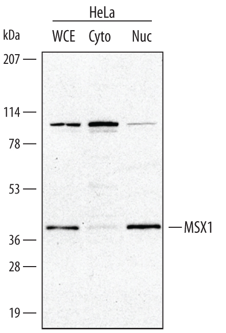
- Detection of Human MSX1 by Western Blot. Western blot shows lysates of HeLa human cervical epithelial carcinoma cell line. Gels were loaded with 30 μg of whole cell lysate (WCL), 20 μg of cytoplasmic (Cyto), and 10 μg of nuclear extracts (Nuc). PVDF membrane was probed with 0.1 µg/mL Goat Anti-Human/Mouse MSX1 Antigen Affinity-purified Polyclonal Antibody (Catalog # AF5045) followed by HRP-conjugated Anti-Goat IgG Secondary Antibody (Catalog # HAF017). A specific band for MSX1 was detected at approximately 40 kDa (as indicated). This experiment was conducted under reducing conditions and using Immunoblot Buffer Group 1.
Supportive validation
- Submitted by
- R&D Systems (provider)
- Main image
- Experimental details
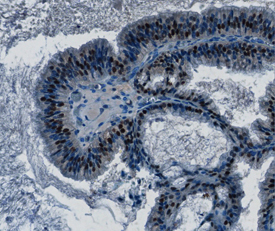
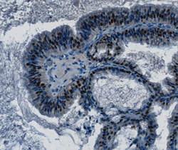
- MSX1 in Human Ovarian Cancer Tissue. MSX1 was detected in immersion fixed paraffin-embedded sections of human ovarian cancer tissue using Goat Anti-Human/Mouse MSX1 Antigen Affinity-purified Polyclonal Antibody (Catalog # AF5045) at 0.3, 1.0 and 3.0 µg/mL overnight at 4 °C. Tissue was stained using the Anti-Goat HRP-DAB Cell & Tissue Staining Kit (brown; Catalog # CTS008) and counterstained with hematoxylin (blue). Specific staining was localized to nuclei. View our protocol for Chromogenic IHC Staining of Paraffin-embedded Tissue Sections.