Explore
 Validate
 Learn
Learn Western blot
 Immunohistochemistry
Antibody data
- Antibody Data
- Antigen structure
- References [4]
- Comments [0]
- Validations
- Immunohistochemistry [1]
Submit
Validation data
Reference
Comment
Report error
- Product number
- HPA018871 - Provider product page

- Provider
- Atlas Antibodies
- Proper citation
- Atlas Antibodies Cat#HPA018871, RRID:AB_1856724
- Product name
- Anti-SERPINB1
- Antibody type
- Polyclonal
- Description
- Polyclonal Antibody against Human SERPINB1, Gene description: serpin peptidase inhibitor, clade B (ovalbumin), member 1, Alternative Gene Names: anti-elastase, EI, ELANH2, PI2, Validated applications: WB, IHC, Uniprot ID: P30740, Storage: Store at +4°C for short term storage. Long time storage is recommended at -20°C.
- Reactivity
- Human
- Host
- Rabbit
- Conjugate
- Unconjugated
- Isotype
- IgG
- Vial size
- 100 µl
- Concentration
- 0.2 mg/ml
- Storage
- Store at +4°C for short term storage. Long time storage is recommended at -20°C.
- Handling
- The antibody solution should be gently mixed before use.
Submitted references Multi-omics analysis of the cervical epithelial integrity of women using depot medroxyprogesterone acetate
A High-throughput Bead-based Affinity Assay Enables Analysis of Genital Protein Signatures in Women At Risk of HIV Infection
SERPINB1 expression is predictive for sensitivity and outcome of cisplatin-based chemotherapy in melanoma
Proteomic analysis reveals heat shock protein 70 has a key role in polycythemia Vera
Tachedjian G, Bradley F, Franzén Boger M, Kaldhusdal V, Åhlberg A, Edfeldt G, Lajoie J, Bergström S, Omollo K, Damdimopoulos A, Czarnewski P, Månberg A, Oyugi J, Kimani J, Nilsson P, Fowke K, Tjernlund A, Broliden K
PLOS Pathogens 2022;18(5):e1010494
PLOS Pathogens 2022;18(5):e1010494
A High-throughput Bead-based Affinity Assay Enables Analysis of Genital Protein Signatures in Women At Risk of HIV Infection
Månberg A, Bradley F, Qundos U, Guthrie B, Birse K, Noël-Romas L, Lindskog C, Bosire R, Kiarie J, Farquhar C, Burgener A, Nilsson P, Broliden K
Molecular & Cellular Proteomics 2019;18(3):461-476
Molecular & Cellular Proteomics 2019;18(3):461-476
SERPINB1 expression is predictive for sensitivity and outcome of cisplatin-based chemotherapy in melanoma
Willmes C, Kumar R, Becker J, Fried I, Rachakonda P, Poppe L, Hesbacher S, Schadendorf D, Sucker A, Schrama D, Ugurel S
Oncotarget 2016;7(9):10117-10132
Oncotarget 2016;7(9):10117-10132
Proteomic analysis reveals heat shock protein 70 has a key role in polycythemia Vera
Gallardo M, Barrio S, Fernandez M, Paradela A, Arenas A, Toldos O, Ayala R, Albizua E, Jimenez A, Redondo S, Garcia-Martin R, Gilsanz F, Albar J, Martinez-Lopez J
Molecular Cancer 2013;12(1):142
Molecular Cancer 2013;12(1):142
No comments: Submit comment
Supportive validation
- Submitted by
- Atlas Antibodies (provider)
- Enhanced method
- Orthogonal validation
- Main image
- Experimental details
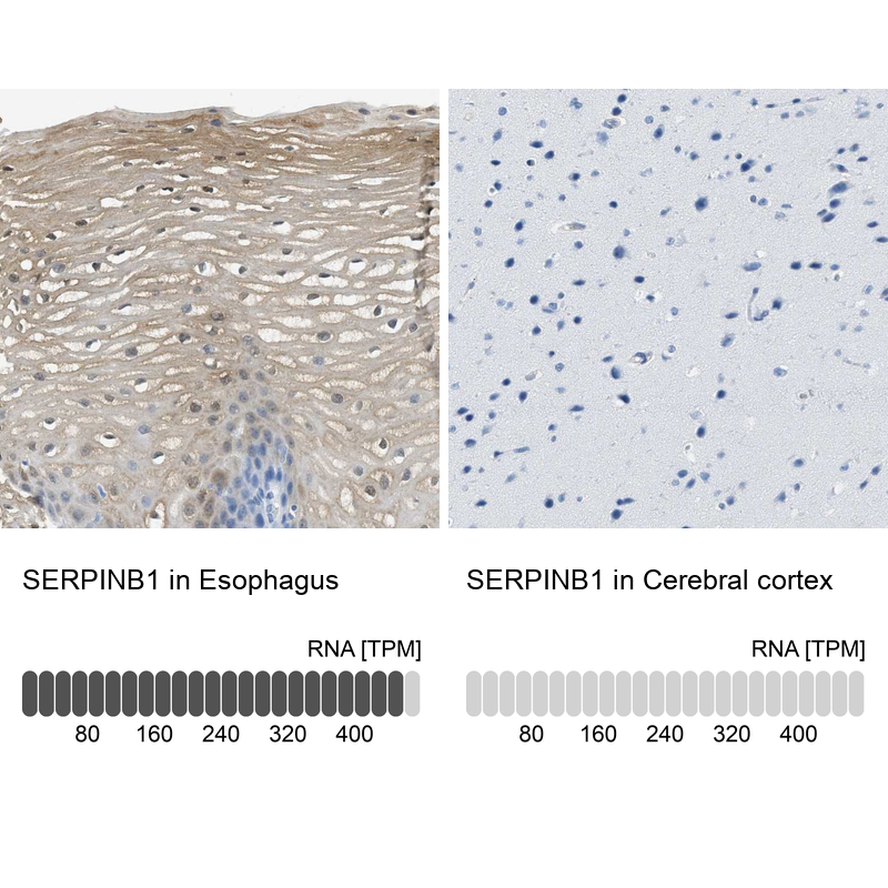
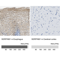
- Immunohistochemistry analysis in human esophagus and cerebral cortex tissues using Anti-SERPINB1 antibody. Corresponding SERPINB1 RNA-seq data are presented for the same tissues.
- Sample type
- Human
- Protocol
- Protocol