 Explore
 Validate
 Learn
Learn702554
antibody from Invitrogen Antibodies
Targeting: FOXG1
BF1, FKH2, FKHL1, FKHL2, FKHL3, FKHL4, FOXG1A, FOXG1B, FOXG1C, HBF-3, HFK1, HFK2, HFK3, QIN
 Western blot
Antibody data
- Antibody Data
- Antigen structure
- References [0]
- Comments [0]
- Validations
- Western blot [2]
- Immunocytochemistry [1]
Submit
Validation data
Reference
Comment
Report error
- Product number
- 702554 - Provider product page

- Provider
- Invitrogen Antibodies
- Product name
- FOXG1 Recombinant Rabbit Monoclonal Antibody (2H7L2)
- Antibody type
- Monoclonal
- Antigen
- Synthetic peptide
- Reactivity
- Human, Mouse
- Host
- Rabbit
- Isotype
- IgG
- Antibody clone number
- 2H7L2
- Vial size
- 100 µg
- Concentration
- 0.5 mg/mL
- Storage
- Store at 4°C short term. For long term storage, store at -20°C, avoiding freeze/thaw cycles.
No comments: Submit comment
Supportive validation
- Submitted by
- Invitrogen Antibodies (provider)
- Main image
- Experimental details
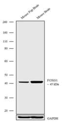
- Western blot analysis was performed on Tissue extracts (30 µg lysate) of Mouse pup Brain (Lane 1) and Mouse Brain (Lane 2). The blots were probed with Anti-FOXG1 Recombinant Rabbit Monoclonal Antibody (Product # 702554, 1-2 µg/mL) and detected by chemiluminescence using Goat anti-Rabbit IgG (H+L) Superclonal™ Secondary Antibody, HRP conjugate (Product # A27036, 0.4 µg/mL, 1:2500 dilution). A 45 kDa band corresponding to FOXG1 was observed across the tissues tested. Known quantity of protein samples were electrophoresed using Novex® NuPAGE® 4-12% Bis-Tris gel (Product # NP0321BOX), XCell SureLock™ Electrophoresis System (Product # EI0002) and Novex® Sharp Pre-Stained Protein Standard (Product # LC5800). Resolved proteins were then transferred onto a nitrocellulose membrane with iBlot® Dry Blotting System (Product # IB21001). The membrane was probed with the relevant primary and secondary Antibody following blocking with 5% skimmed milk. Chemiluminescent detection was performed using Pierce™ ECL Western blotting Substrate (Product # 32106).

- Submitted by
- Invitrogen Antibodies (provider)
- Main image
- Experimental details
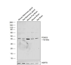
- Western blot was performed using Anti-FOXG1 Recombinant Rabbit Monoclonal Antibody (2H7L2) (Product # 702554) and a 42 kDa band corresponding to FOXG1 was observed across tissues tested. Tissue extracts (30 µg lysate) of Mouse Mammary gland (Lane 1), Rat Mammary gland (Lane 2), Mouse Pup Brain (Lane 3), Mouse Brain (Lane 4), Rat Brain (Lane 5) were electrophoresed using NuPAGE™ 4-12% Bis-Tris Protein Gel (Product # NP0321BOX). Resolved proteins were then transferred onto a Nitrocellulose membrane (Product # IB23001) by iBlot® 2 Dry Blotting System (Product # IB21001). The blot was probed with the primary antibody (2 ug/ml) and detected by chemiluminescence with Goat anti-Rabbit IgG (H+L) Superclonal™ Recombinant Secondary Antibody, HRP (Product # A27036,1:10000 dilution) using the iBright FL 1000 (Product # A32752). Chemiluminescent detection was performed using SuperSignal™ West Dura Extended Duration Substrate (Product # 34076).

Supportive validation
- Submitted by
- Invitrogen Antibodies (provider)
- Main image

- Experimental details
- For immunofluorescence analysis, U-87 MG cells were fixed and permeabilized for detection of endogenous FOXG1 using Anti-FOXG1 Recombinant Rabbit Monoclonal Antibody (Product # 702554, 2 µg/mL) and labeled with Goat anti-Rabbit IgG (H+L) Superclonal™ Secondary Antibody, Alexa Fluor® 488 conjugate (Product # A27034, 1:2000). Panel a) shows representative cells that were stained for detection and localization of FOXG1 protein (green), Panel b) is stained for nuclei (blue) using SlowFade® Gold Antifade Mountant with DAPI (Product # S36938). Panel c) represents cytoskeletal F-actin staining using Rhodamine Phalloidin (Product # R415, 1:300). Panel d) is a composite image of Panels a, b and c clearly demonstrating nuclear localization of FOXG1 in U-87 mg glioblastoma cells which is positive for FOXG1 (Seoane et al., 2004). Panel e)represents control cells with no primary antibody to assess background panel f) shows no signal in SH-SY5Y cells which is negative for FOXG1.The images were captured at 60X magnification.
