 Explore
 Validate
 Learn
LearnPA5-26794
antibody from Invitrogen Antibodies
Targeting: FOXG1
BF1, FKH2, FKHL1, FKHL2, FKHL3, FKHL4, FOXG1A, FOXG1B, FOXG1C, HBF-3, HFK1, HFK2, HFK3, QIN
 Western blot
Antibody data
- Antibody Data
- Antigen structure
- References [2]
- Comments [0]
- Validations
- Western blot [3]
- Immunocytochemistry [2]
- Flow cytometry [2]
- Other assay [5]
Submit
Validation data
Reference
Comment
Report error
- Product number
- PA5-26794 - Provider product page

- Provider
- Invitrogen Antibodies
- Product name
- FOXG1 Polyclonal Antibody
- Antibody type
- Polyclonal
- Antigen
- Synthetic peptide
- Description
- This antibody is predicted to react with chicken, canine, rat and Xenopus based on sequence homology.
- Reactivity
- Human, Mouse
- Host
- Rabbit
- Isotype
- IgG
- Vial size
- 400 μL
- Concentration
- 2 mg/mL
- Storage
- Store at 4°C short term. For long term storage, store at -20°C, avoiding freeze/thaw cycles.
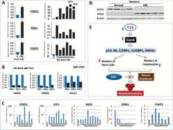
Submitted references FXR-Gankyrin axis is involved in development of pediatric liver cancer.
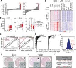
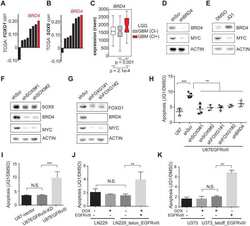
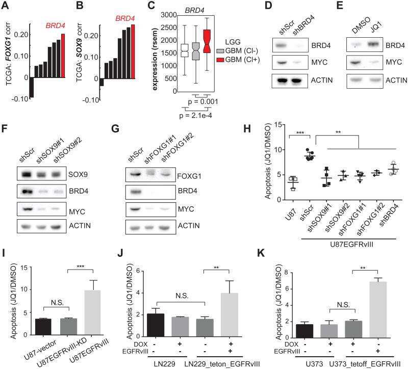
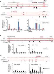
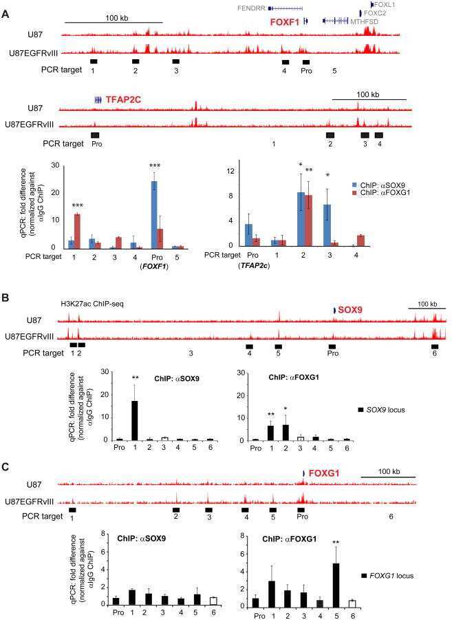
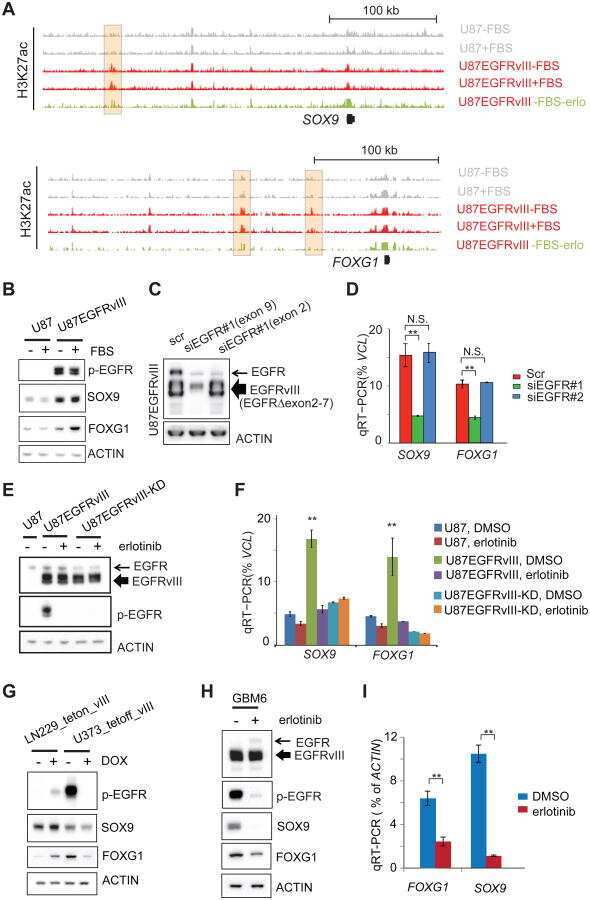
EGFR Mutation Promotes Glioblastoma through Epigenome and Transcription Factor Network Remodeling.
Valanejad L, Lewis K, Wright M, Jiang Y, D'Souza A, Karns R, Sheridan R, Gupta A, Bove K, Witte D, Geller J, Tiao G, Nelson DL, Timchenko L, Timchenko N
Carcinogenesis 2017 Jul 1;38(7):738-747
Carcinogenesis 2017 Jul 1;38(7):738-747
EGFR Mutation Promotes Glioblastoma through Epigenome and Transcription Factor Network Remodeling.
Liu F, Hon GC, Villa GR, Turner KM, Ikegami S, Yang H, Ye Z, Li B, Kuan S, Lee AY, Zanca C, Wei B, Lucey G, Jenkins D, Zhang W, Barr CL, Furnari FB, Cloughesy TF, Yong WH, Gahman TC, Shiau AK, Cavenee WK, Ren B, Mischel PS
Molecular cell 2015 Oct 15;60(2):307-18
Molecular cell 2015 Oct 15;60(2):307-18
No comments: Submit comment
Supportive validation
- Submitted by
- Invitrogen Antibodies (provider)
- Main image

- Experimental details
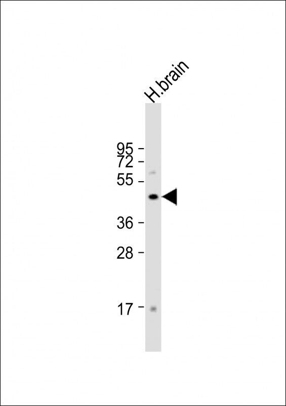
- Western blot analysis of FOXG1 in Human brain lysate. Samples were incubated with FOXG1 polyclonal antibody (Product # PA5-26794) using a dilution of 1:1,000 followed by Goat Anti-Rabbit IgG, (H+L), Peroxidase conjugated at a dilution of 1:10,000. Lysates/proteins: 20 µg per lane. Predicted band size: 52 kDa. Blocking/Dilution buffer: 5% NFDM/TBST.
- Submitted by
- Invitrogen Antibodies (provider)
- Main image
- Experimental details
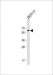
- Western blot analysis of FOXG1 in 293T/17 whole cell lysate. Samples were incubated with FOXG1 polyclonal antibody (Product # PA5-26794) using a dilution of 1:1,000 followed by Goat Anti-Rabbit IgG, (H+L), Peroxidase conjugated at a dilution of 1:15,000. Lysates/proteins: 20 µg per lane.

- Submitted by
- Invitrogen Antibodies (provider)
- Main image

- Experimental details
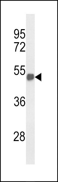
- Western blot analysis of FOXG1 in mouse brain tissue lysates. Samples were incubated with FOXG1 polyclonal antibody (Product # PA5-26794). Lysates: 35 µg/lane. FOXG1 (arrow).
Supportive validation
- Submitted by
- Invitrogen Antibodies (provider)
- Main image

- Experimental details
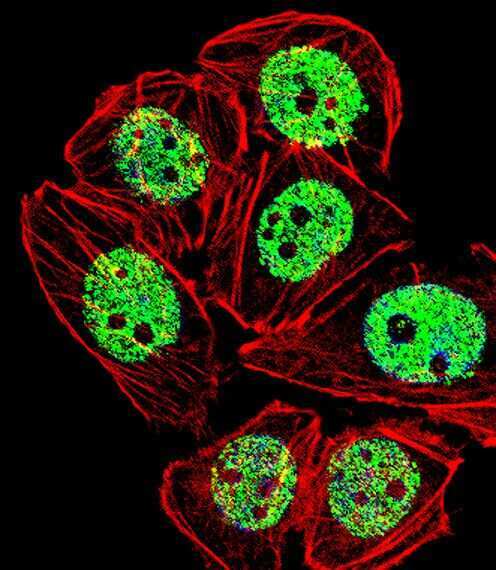
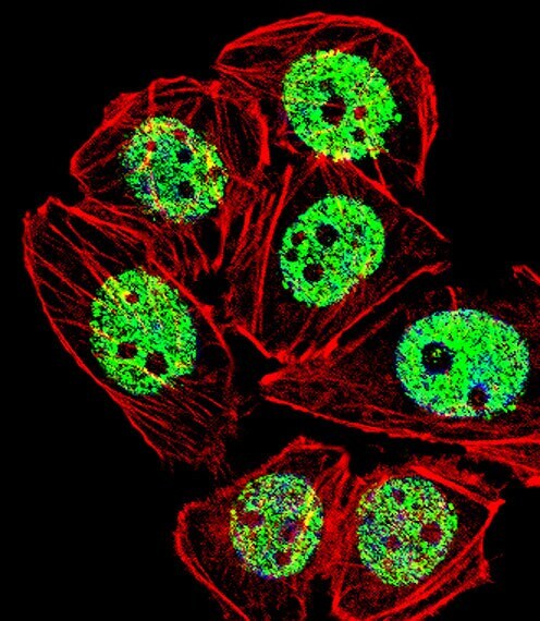
- Immunofluorescent analysis of A549 cells using a FOXG1 polyclonal antibody (Product # PA5-26794). A549 cells were fixed with 4% PFA (20 min), permeabilized with Triton X-100 (0.1%, 10 min), then incubated with a FOXG1 polyclonal antibody (Product # PA5-26794) (1:25, 1 hr at 37°C). Primary antibody was detected with fluor-conjugated donkey anti-rabbit secondary antibody (green) at 1:400 dilution for 50 min at 37°C). Actin filaments have been labeled with dye-conjugated phalloidin (red). Nuclei were counterstained with DAPI (blue) (10 µg/mL, 10 min).
- Submitted by
- Invitrogen Antibodies (provider)
- Main image

- Experimental details
- Immunocytochemistry analysis of FOXG1 in A549 cells. Samples were incubated with FOXG1 polyclonal antibody (Product # PA5-26794) using a dilution of 1:25 for 1 h at 37°C followed by Alexa Fluor® 488 conjugated donkey anti-rabbit at a dilution of 1:400 for 50 min at 37°C. Cells were fixed with 4% PFA (20 min) and permeabilized with Triton X-100 (0.1%, 10 min). Cytoplasmic actin was counterstained with Alexa Fluor® 555 (red) conjugated Phalloidin (7 units/mL, 1 h at 37°C). Nuclei were counterstained with DAPI (blue) (10 µg/mL, 10 min). FOXG1 immunoreactivity is localized to Nucleus significantly.
Supportive validation
- Submitted by
- Invitrogen Antibodies (provider)
- Main image
- Experimental details
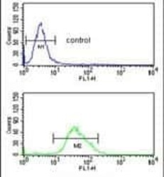
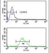
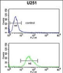
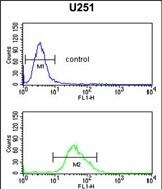
- Flow cytometry analysis of U251 cells using a FOXG1 polyclonal antibody (Product # PA5-26794) (bottom) compared to a negative control cell (top) at a dilution of 1:10-50, followed by a FITC-conjugated goat anti-rabbit antibody
- Submitted by
- Invitrogen Antibodies (provider)
- Main image
- Experimental details
- Flow cytometry of FOXG1 in U251 cells (bottom histogram). Samples were incubated with FOXG1 polyclonal antibody (Product # PA5-26794) followed by FITC-conjugated goat-anti-rabbit secondary antibody. Negative control cell (top histogram).
Supportive validation
- Submitted by
- Invitrogen Antibodies (provider)
- Main image
- Experimental details
- NULL

- Submitted by
- Invitrogen Antibodies (provider)
- Main image
- Experimental details
- NULL
- Submitted by
- Invitrogen Antibodies (provider)
- Main image
- Experimental details
- NULL
- Submitted by
- Invitrogen Antibodies (provider)
- Main image
- Experimental details
- NULL
- Submitted by
- Invitrogen Antibodies (provider)
- Main image
- Experimental details
- NULL
