 Explore
 Validate
 Learn
Learn Western blot
Antibody data
- Antibody Data
- Antigen structure
- References [5]
- Comments [0]
- Validations
- Western blot [3]
- Immunocytochemistry [4]
- Other assay [3]
Submit
Validation data
Reference
Comment
Report error
- Product number
- PA1-333 - Provider product page

- Provider
- Invitrogen Antibodies
- Product name
- LXR beta Polyclonal Antibody
- Antibody type
- Polyclonal
- Antigen
- Synthetic peptide
- Description
- PA1-333 detects LXR beta in human, rat and mouse samples. PA1-333 has been successfully used in Western blot and IF procedures. By Western blot, this antibody detects an ~51 kDa protein representing LXR beta in rat and mouse liver samples. The PA1-333 immunogen is a synthetic peptide corresponding to residues R(113) Y A C R G S G T C Q M D A(126) of rat LXR beta. The PA1-333 immunizing peptide (Cat. # PEP-271) is available for use in neutralization and control experiments.
- Reactivity
- Human, Mouse, Rat
- Host
- Rabbit
- Isotype
- IgG
- Vial size
- 100 μg
- Concentration
- 1 mg/mL
- Storage
- -20°C, Avoid Freeze/Thaw Cycles
Submitted references Liver X receptor β is required for the survival of single-positive thymocytes by regulating IL-7Rα expression.
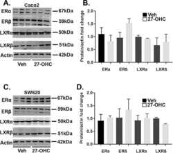
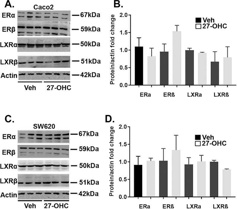
27-hydroxycholesterol decreases cell proliferation in colon cancer cell lines.
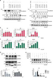
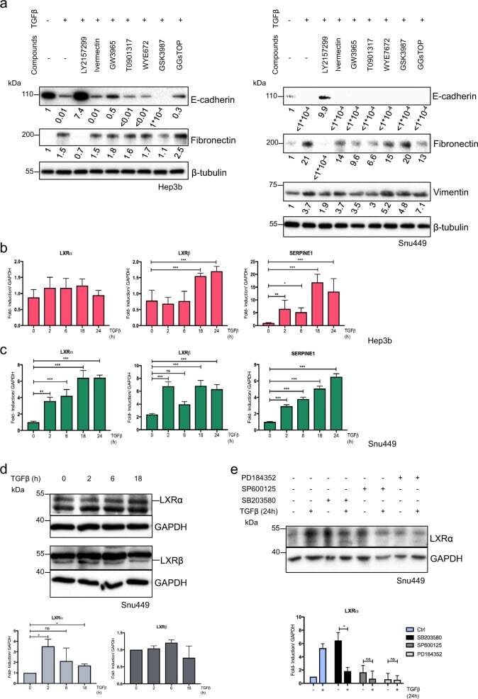
Snail mediates crosstalk between TGFβ and LXRα in hepatocellular carcinoma.
Macrophage-derived IL-1β/NF-κB signaling mediates parenteral nutrition-associated cholestasis.
ALOX5AP Overexpression in Adipose Tissue Leads to LXA4 Production and Protection Against Diet-Induced Obesity and Insulin Resistance.
Huang H, Wu X, Meng D, Feng Y, Zhou L, Liu Z, Tang S, Li X, Cao Y, He H, Xie Z, Zhang J, Chen Y, Zhao T, Wu Y, Zhou X
Cellular & molecular immunology 2021 Aug;18(8):1969-1980
Cellular & molecular immunology 2021 Aug;18(8):1969-1980
27-hydroxycholesterol decreases cell proliferation in colon cancer cell lines.
Warns J, Marwarha G, Freking N, Ghribi O
Biochimie 2018 Oct;153:171-180
Biochimie 2018 Oct;153:171-180
Snail mediates crosstalk between TGFβ and LXRα in hepatocellular carcinoma.
Bellomo C, Caja L, Fabregat I, Mikulits W, Kardassis D, Heldin CH, Moustakas A
Cell death and differentiation 2018 May;25(5):885-903
Cell death and differentiation 2018 May;25(5):885-903
Macrophage-derived IL-1β/NF-κB signaling mediates parenteral nutrition-associated cholestasis.
El Kasmi KC, Vue PM, Anderson AL, Devereaux MW, Ghosh S, Balasubramaniyan N, Fillon SA, Dahrenmoeller C, Allawzi A, Woods C, McKenna S, Wright CJ, Johnson L, D'Alessandro A, Reisz JA, Nozik-Grayck E, Suchy FJ, Sokol RJ
Nature communications 2018 Apr 11;9(1):1393
Nature communications 2018 Apr 11;9(1):1393
ALOX5AP Overexpression in Adipose Tissue Leads to LXA4 Production and Protection Against Diet-Induced Obesity and Insulin Resistance.
Elias I, Ferré T, Vilà L, Muñoz S, Casellas A, Garcia M, Molas M, Agudo J, Roca C, Ruberte J, Bosch F, Franckhauser S
Diabetes 2016 Aug;65(8):2139-50
Diabetes 2016 Aug;65(8):2139-50
No comments: Submit comment
Supportive validation
- Submitted by
- Invitrogen Antibodies (provider)
- Main image

- Experimental details
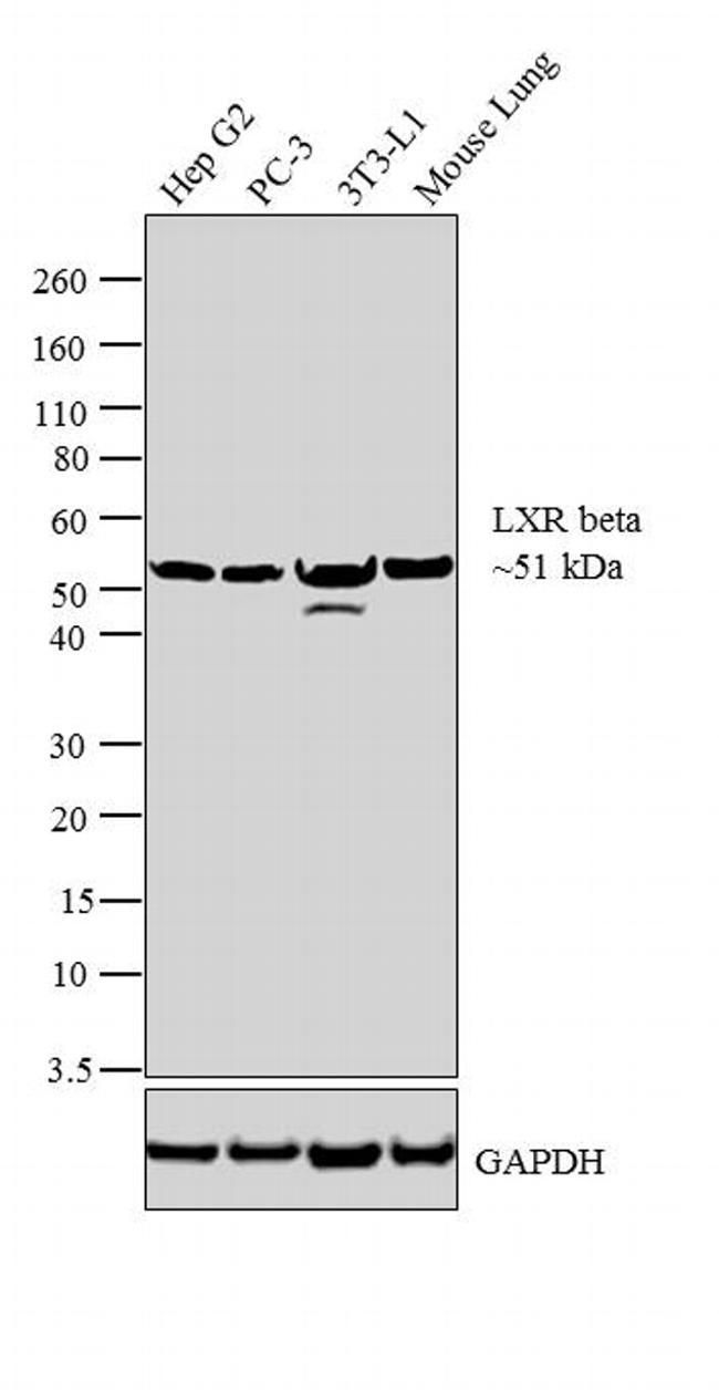
- Western blot analysis was performed on nuclear enriched extracts (30µg lysate) of Hep G2 (Lane 1), PC-3 (Lane 2), 3T3-L1 (Lane 3) and tissue extract of Mouse Lung (Lane 4). The blot was probed with Rabbit Anti-LXR beta Polyclonal Antibody (Product # PA1-333, 2µg/mL) and detected by chemiluminescence using Goat anti-Rabbit IgG (Heavy Chain) Superclonal™ Secondary Antibody, HRP conjugate (Product # A27036, 0.25µg/mL, 1:4000 dilution). A 51 kDa band corresponding to LXR beta was observed across the cell lines and tissue tested. Known quantity of protein samples were electrophoresed using Novex® NuPAGE® 4-12 % Bis-Tris gel (Product # NP0321BOX), XCell SureLock™ Electrophoresis System (Product # EI0002) and Novex® Sharp Pre-Stained Protein Standard (Product # LC5800). Resolved proteins were then transferred onto a nitrocellulose membrane with iBlot® 2 Dry Blotting System (Product # IB21001). The membrane was probed with the relevant primary and secondary Antibody using iBind™ Flex Western Starter Kit (Product # SLF2000S). Chemiluminescent detection was performed using Pierce™ ECL Western Blotting Substrate (Product # 32106).
- Submitted by
- Invitrogen Antibodies (provider)
- Main image
- Experimental details
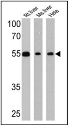
- Western blot analysis of LXR beta was performed by loading 25 µg of rat liver (Lane 1), mouse liver (Lane 2) and Hela (Lane 3) cell lysates onto an SDS polyacrylamide gel. Proteins were transferred to a PVDF membrane and blocked at 4ºC overnight. The membrane was probed with a LXR beta polyclonal antibody (Product # PA1-333) at a dilution of 1:1000 overnight at 4°C, washed in TBST, and probed with an HRP-conjugated secondary antibody for 1 hr at room temperature in the dark. Chemiluminescent detection was performed using Pierce ECL Plus Western Blotting Substrate (Product # 32132). Results show a band at approx. 55 kDa.

- Submitted by
- Invitrogen Antibodies (provider)
- Main image
- Experimental details
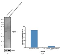
- CRISPR-Cas9 mediated genome editing of LXR beta (as confirmed by next generation sequencing) was achieved by using LentiArray™ Lentiviral sgRNA (Product # A32042, AssayID CRISPR765314_LV) and LentiArray Cas9 Lentivirus (Product # A32064). Western blot analysis of LXR beta was performed by loading 30 µg of Hep G2 CAS9 (Lane 1) and Hep G2 Cas9 cells transduced with LXR beta lentiviral sgRNA (Lane 2) modified whole cell extracts. The samples were electrophoresed using NuPAGE™ Novex™ 4-12% Bis-Tris Protein Gel (Product # NP0321BOX). Resolved proteins were then transferred onto a nitrocellulose membrane (Product # IB23001) by iBlot® 2 Dry Blotting System (Product # IB21001). The blot was probed with LXR beta Polyclonal Antibody (Product # PA1-333, 1:500) and detected by Goat anti-Rabbit IgG (H+L) Superclonal™ Recombinant Secondary Antibody, HRP (Product # A27036 1:10,000) using iBright™ FL1500 (Product # A44115). Chemiluminescent detection was performed using SuperSignal™ West Atto Ultimate Sensitivity Substrate (Product # A38556). A reduced signal in sgRNA transduced cells using the LentiArray™ CRISPR product line confirms that antibody is specific to LXR beta.

Supportive validation
- Submitted by
- Invitrogen Antibodies (provider)
- Main image

- Experimental details
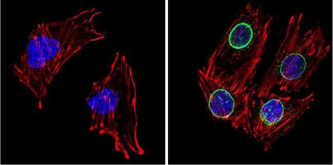
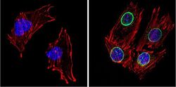
- Immunofluorescent analysis of LXR beta (green) showing positive staining in the nuclear envelope of C2C12 cells (right) compared with a negative control in the absence of primary antibody (left). Formalin-fixed cells were permeabilized with 0.1% Triton X-100 in TBS for 5-10 minutes, blocked with 3% BSA-PBS for 30 minutes at room temperature and probed with an LXR polyclonal antibody (Product # PA1-333) in 3% BSA-PBS at a dilution of 1:200 and incubated overnight at 4 ºC in a humidified chamber. Cells were washed with PBST and incubated with a DyLight 488-conjugated goat-anti-rabbit IgG secondary antibody in PBS at room temperature in the dark. F-actin (red) was stained with a fluorescent red phalloidin and nuclei (blue) were stained with DAPI for 5-10 minutes in the dark. Images were taken at a magnification of 60x.
- Submitted by
- Invitrogen Antibodies (provider)
- Main image
- Experimental details
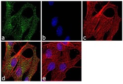
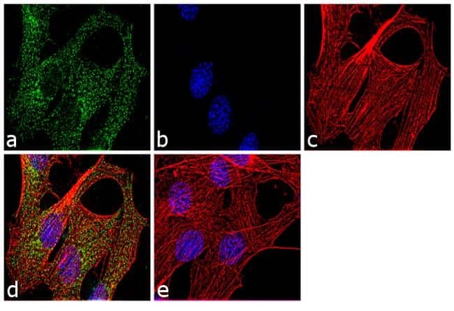
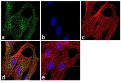
- Immunofluorescence analysis of LXR beta was performed using 70% confluent log phase C2C12 cells. The cells were fixed with 4% paraformaldehyde for 10 minutes, permeabilized with 0.1% Triton™ X-100 for 10 minutes, and blocked with 1% BSA for 1 hour at room temperature. The cells were labeled with LXR beta Rabbit Polyclonal Antibody (Product # PA1-333) at 2µg/mL in 0.1% BSA and incubated for 3 hours at room temperature and then labeled with Goat anti-Rabbit IgG (H+L) Superclonal™ Secondary Antibody, Alexa Fluor® 488 conjugate (Product # A27034) at a dilution of 1:2000 for 45 minutes at room temperature (Panel a: green). Nuclei (Panel b: blue) were stained with SlowFade® Gold Antifade Mountant with DAPI (Product # S36938). F-actin (Panel c: red) was stained with Rhodamine Phalloidin (Product # R415, 1:300). Panel d represents the merged image showing cytoplasmic and nuclear localization. Panel e shows the no primary antibody control. The images were captured at 60X magnification.
- Submitted by
- Invitrogen Antibodies (provider)
- Main image
- Experimental details
- Immunofluorescent analysis of LXR beta (green) showing positive staining in the nuclear envelope of C2C12 cells (right) compared with a negative control in the absence of primary antibody (left). Formalin-fixed cells were permeabilized with 0.1% Triton X-100 in TBS for 5-10 minutes, blocked with 3% BSA-PBS for 30 minutes at room temperature and probed with an LXR polyclonal antibody (Product # PA1-333) in 3% BSA-PBS at a dilution of 1:200 and incubated overnight at 4 ºC in a humidified chamber. Cells were washed with PBST and incubated with a DyLight 488-conjugated goat-anti-rabbit IgG secondary antibody in PBS at room temperature in the dark. F-actin (red) was stained with a fluorescent red phalloidin and nuclei (blue) were stained with DAPI for 5-10 minutes in the dark. Images were taken at a magnification of 60x.

- Submitted by
- Invitrogen Antibodies (provider)
- Main image
- Experimental details
- Immunofluorescence analysis of LXR beta was performed using 70% confluent log phase C2C12 cells. The cells were fixed with 4% paraformaldehyde for 10 minutes, permeabilized with 0.1% Triton™ X-100 for 10 minutes, and blocked with 1% BSA for 1 hour at room temperature. The cells were labeled with LXR beta Rabbit Polyclonal Antibody (Product # PA1-333) at 2µg/mL in 0.1% BSA and incubated for 3 hours at room temperature and then labeled with Goat anti-Rabbit IgG (Heavy Chain) Superclonal™ Secondary Antibody, Alexa Fluor® 488 conjugate (Product # A27034) at a dilution of 1:2000 for 45 minutes at room temperature (Panel a: green). Nuclei (Panel b: blue) were stained with SlowFade® Gold Antifade Mountant with DAPI (Product # S36938). F-actin (Panel c: red) was stained with Rhodamine Phalloidin (Product # R415, 1:300). Panel d represents the merged image showing cytoplasmic and nuclear localization. Panel e shows the no primary antibody control. The images were captured at 60X magnification.

Supportive validation
- Submitted by
- Invitrogen Antibodies (provider)
- Main image
- Experimental details
- NULL
- Submitted by
- Invitrogen Antibodies (provider)
- Main image
- Experimental details
- Fig. 1 TGFbeta regulates LXRalpha expression in the mesenchymal cell line Snu449 a Hep3B (left) and Snu449 (right) cells were treated with the indicated compounds (10 muM) and TGFbeta1 (5 ng/ml) for 24 h. The TGFbeta type I receptor kinase inhibitor LY2157299 was added at a final concentration of 2 muM for 24 h as a potent positive control. The expression of the indicated proteins was analyzed by immunoblotting. Experiments performed in biological duplicate. Densitometric quantification is provided below each lane. Results indicated as
- Submitted by
- Invitrogen Antibodies (provider)
- Main image
- Experimental details
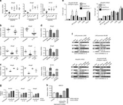
- Fig. 5 IL-1beta induced NF-kappaB signaling suppresses LXR and ABCG5/8 . a Liver mRNA for Abcg5 , Abcg8 , and Nr1h3 in WT chow mice relative to WT DSS/PN, WT PN-only, Ccr2 -/- DSS/PN, Il1r -/- DSS/PN, and Anakinra DSS/PN mice. b , c Liver mRNA for Abcg5 and Abcg8 in WT mice left untreated, or IL-1beta i.p. injected (4 h) (upper panels) and in mice LPS i.p. injected (4 h), with or without prior (16 h) i.p. clodronate-liposome (Clo-lip) injection (bottom panels). * p < 0.05 vs. all other groups using t test (upper panels) or one-way ANOVA and Tukey's correction (lower panels). c Liver mRNA for Abcg5 and Abcg8 in WT mice left untreated, or i.p. LPS injected relative to that Casp1/11 -/- and Il1r -/- mice. d Liver mRNA for Abcg5 , Abcg8 , and Nr1h3 in untreated mouse primary hepatocytes relative to LXR agonist (GW3965, 2 muM, 16 h) and IL-1beta (additional 4 h) treatment. e qPCR from LXRbeta binding site within the ABCG5/8 intergenic region and ChIP for LXRalpha or NF-kappaB p65 and p50 on liver homogenate from WT chow, WT DSS/PN, and Il1r -/- DSS/PN mice. Data represented as fold change over IgG; POLII shown as control. f ChIP from HuH7 cells left untreated (HuH7), or exposed to LXR agonist (GW3965, 2 muM; HuH7 + GW3965) for 16 h with or without additional exposure to IL-1beta for 4 h. Specific antibodies for either LXR (left) binding to LXR promoter (top left) or ABCG8 intergenic region (bottom left) or NF-kappaB p50 and p65 (ri
