Explore
 Validate
 Learn
Learn Western blot
Antibody data
- Antibody Data
- Antigen structure
- References [4]
- Comments [0]
- Validations
- Western blot [2]
- Immunocytochemistry [2]
Submit
Validation data
Reference
Comment
Report error
- Product number
- MA1-811 - Provider product page

- Provider
- Invitrogen Antibodies
- Product name
- RARB Monoclonal Antibody (R beta 28)
- Antibody type
- Monoclonal
- Antigen
- Synthetic peptide
- Description
- MA1-811 detects retinoic acid receptor (RAR) beta from human and mouse tissues. This antibody cross-reacts slightly with RAR alpha when expressed in COS cells, though the level of signal on Western blots is very low relative to that seen with RAR beta. It does not detect RAR alpha or gamma from cell lines or tissue extract. MA1-811 has been successfully used in Western blot procedures. By Western blot, this antibody detects an ~52 kDa protein in rat tissue extract representing RAR beta. Also, by Western blot MA1-811 detects an unidentified ~120 kDa protein which can be competed away with the immunizing peptide. This protein is most notable in human SH-SY5Y and COS cells. Due to the extremely low levels of RAR expressed in native tissues, it is recommended that sensitive detection systems, such as enhanced chemiluminescence, be used. The MA1-811 immunogen is a synthetic peptide corresponding to the C-terminal sequence of mouse RAR beta. Reconstitute with PBS. MA1-811 can be used with blocking peptide PEP-005.
- Reactivity
- Human, Mouse, Rat
- Host
- Mouse
- Isotype
- IgG
- Antibody clone number
- R beta 28
- Vial size
- 50 μg
- Concentration
- Conc. Not Determined
- Storage
- -20°C, Avoid Freeze/Thaw Cycles
Submitted references Downregulation of RAR alpha in mice by antisense transgene leads to a compensatory increase in RAR beta and RAR gamma and development of lymphoma.
Arg269 and Lys220 of retinoic acid receptor-beta are important for the binding of retinoic acid.
Transactivation properties of retinoic acid and retinoid X receptors in mammalian cells and yeast. Correlation with hormone binding and effects of metabolism.
Identification of human, mouse, and rat retinoic acid receptor alpha using monoclonal antibodies.
Manshouri T, Yang Y, Lin H, Stass SA, Glassman AB, Keating MJ, Albitar M
Blood 1997 Apr 1;89(7):2507-15
Blood 1997 Apr 1;89(7):2507-15
Arg269 and Lys220 of retinoic acid receptor-beta are important for the binding of retinoic acid.
Tairis N, Gabriel JL, Gyda M 3rd, Soprano KJ, Soprano DR
The Journal of biological chemistry 1994 Jul 29;269(30):19516-22
The Journal of biological chemistry 1994 Jul 29;269(30):19516-22
Transactivation properties of retinoic acid and retinoid X receptors in mammalian cells and yeast. Correlation with hormone binding and effects of metabolism.
Allegretto EA, McClurg MR, Lazarchik SB, Clemm DL, Kerner SA, Elgort MG, Boehm MF, White SK, Pike JW, Heyman RA
The Journal of biological chemistry 1993 Dec 15;268(35):26625-33
The Journal of biological chemistry 1993 Dec 15;268(35):26625-33
Identification of human, mouse, and rat retinoic acid receptor alpha using monoclonal antibodies.
Ali M, Torian BE, Vedeckis WV
Biochemical and biophysical research communications 1992 Feb 14;182(3):1032-9
Biochemical and biophysical research communications 1992 Feb 14;182(3):1032-9
No comments: Submit comment
Supportive validation
- Submitted by
- Invitrogen Antibodies (provider)
- Main image
- Experimental details
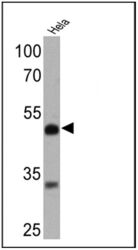
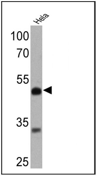
- Western blot analysis of Retinoic Acid Receptor beta was performed by loading 25 µg of Hela cell lysates onto an SDS polyacrylamide gel. Proteins were transferred to a PVDF membrane and blocked at 4ºC overnight. The membrane was probed with a Retinoic Acid Receptor beta monoclonal antibody (Product # MA1-811) at a dilution of 1:1000 overnight at 4°C, washed in TBST, and probed with an HRP-conjugated secondary antibody for 1 hr at room temperature in the dark. Chemiluminescent detection was performed using Pierce ECL Plus Western Blotting Substrate (Product # 32132). Results show a band at ~52 kDa.
- Submitted by
- Invitrogen Antibodies (provider)
- Main image
- Experimental details
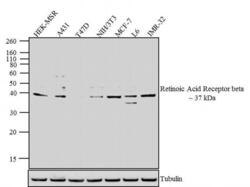
- Western blot analysis was performed on whole cell extracts (30 µg lysate) of HEK-MSR (lane 1), A431 (lane 2), T47D (lane 3), NIH/3T3 (lane 4), MCF-7 (lane 5), L6 (lane 6) and IMR-32 (lane 7). The blots were probed with Anti-Retinoic Acid Receptor beta Mouse Monoclonal Antibody (Product # MA1-811, 1:500-1:2000 dilution) and detected by chemiluminescence Goat anti-Mouse IgG (H+L) Secondary Antibody, HRP conjugate (Product # 62-6520, 1:4000 dilution). A 37 kDa corresponding to Retinoic Acid Receptor beta was observed across cell lines tested expect T47D. Known quantity of protein samples were electrophoresed using Novex® NuPAGE® 10 % Bis-Tris gel (Product # NP0301BOX), XCell SureLock™ Electrophoresis System (Product # EI0002) and Novex® Sharp Pre-Stained Protein Standard (Product # LC5800). Resolved proteins were then transferred onto a nitrocellulose membrane with iBlot® 2 Dry Blotting System (Product # IB21001). The membrane was probed with the relevant primary and secondary Antibody using iBind™ Flex Western Starter Kit (Product # SLF2000S). Chemiluminescent detection was performed using Pierce™ ECL Western Blotting Substrate (Product # 32106).

Supportive validation
- Submitted by
- Invitrogen Antibodies (provider)
- Main image
- Experimental details
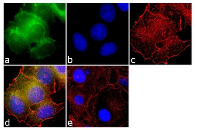
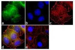
- Immunofluorescence analysis of Retinoic Acid Receptor beta was done on 70% confluent log phase MCF-7 cells. The cells were fixed with 4% paraformaldehyde for 10 minutes, permeabilized with 0.1% Triton™ X-100 for 10 minutes, and blocked with 1% BSA for 1 hour at room temperature. The cells were labeled with Retinoic Acid Receptor beta (R beta 28) Mouse Monoclonal Antibody (Product # MA1-811) at 1:250 dilution in 0.1% BSA and incubated for 3 hours at room temperature and then labeled with Goat anti-Mouse IgG (H+L) Superclonal™ Secondary Antibody, Alexa Fluor® 488 conjugate (Product # A28175) at a dilution of 1:2000 for 45 minutes at room temperature (Panel a: green). Nuclei (Panel b: blue) were stained with SlowFade® Gold Antifade Mountant with DAPI (Product # S36938). F-actin (Panel c: red) was stained with Rhodamine Phalloidin (Product # R415, 1:300). Panel d is a merged image showing cytoplasmic and nuclear localization. Panel e is a no primary antibody control. The images were captured at 60X magnification.

- Submitted by
- Invitrogen Antibodies (provider)
- Main image

- Experimental details
- Immunofluorescence analysis of Retinoic Acid Receptor beta was done on 70% confluent log phase MCF-7 cells. The cells were fixed with 4% paraformaldehyde for 10 minutes, permeabilized with 0.1% Triton™ X-100 for 10 minutes, and blocked with 1% BSA for 1 hour at room temperature. The cells were labeled with Retinoic Acid Receptor beta (R beta 28) Mouse Monoclonal Antibody (Product # MA1-811) at 1:250 dilution in 0.1% BSA and incubated for 3 hours at room temperature and then labeled with Goat anti-Mouse IgG (H+L) Superclonal™ Secondary Antibody, Alexa Fluor® 488 conjugate (Product # A28175) at a dilution of 1:2000 for 45 minutes at room temperature (Panel a: green). Nuclei (Panel b: blue) were stained with SlowFade® Gold Antifade Mountant with DAPI (Product # S36938). F-actin (Panel c: red) was stained with Rhodamine Phalloidin (Product # R415, 1:300). Panel d is a merged image showing cytoplasmic and nuclear localization. Panel e is a no primary antibody control. The images were captured at 60X magnification.