 Explore
 Validate
 Learn
Learn Flow cytometry
Antibody data
- Antibody Data
- Antigen structure
- References [7]
- Comments [0]
- Validations
- Flow cytometry [1]
- Other assay [2]
Submit
Validation data
Reference
Comment
Report error
- Product number
- 12-9816-80 - Provider product page

- Provider
- Invitrogen Antibodies
- Product name
- RUNX1 Monoclonal Antibody (RXDMC), PE, eBioscience™
- Antibody type
- Monoclonal
- Antigen
- Other
- Description
- Description: This RXDMC monoclonal antibody reacts with human and mouse Runx1, which is also known as AML1. This 50-kDa transcription factor is expressed in hematopoietic cells such as myeloid, T, and B cells, but not erythroid cells. Furthermore, Runx1 can be detected in nearly all non-hematopoietic tissues except the brain and heart. Runx1 is involved in several stages of T cell development, including the CD4-CD8- double negative (DN) to CD4+CD8+ double positive (DP) transition and commitment to the CD4 lineage. In addition to its role in thymocyte development, Runx1 has also been implicated in Th2 differentiation and immune homeostasis via direct interaction with Foxp3 in CD4+CD25+ regulatory T cells. Runx1 also affects Th2 differentiation by negatively regulating Gata-3 expression. Finally, the AML1 gene, which encodes Runx1, is a frequent target of translocations in acute myeloid leukemia. In humans, alternative splicing results in the generation of eleven isoforms. In mice, five isoforms exist as a result of alternative splicing; isoform 4 (aka AML1-C) is the most highly expressed in hematopoietic cells. Sequence analysis indicates that the RXDMC antibody recognizes isoform 4 in mouse and all human isoforms except AML-1FA, AML-1FB, and AML-1FC. Applications Reported: This RXDMC antibody has been reported for use in intracellular staining followed by flow cytometric analysis. Applications Tested: This RXDMC antibody has been tested by intracellular staining and flow cytometric analysis of mouse thymocytes using the Foxp3/Transcription Factor Staining Buffer Set (Product # 00-5523-00) and protocol. This can be used at less than or equal to 1 µg per test. A test is defined as the amount (µg) of antibody that will stain a cell sample in a final volume of 100 µL. Cell number should be determined empirically but can range from 10^5 to 10^8 cells/test. It is recommended that the antibody be carefully titrated for optimal performance in the assay of interest. Excitation: 488-561 nm; Emission: 578 nm; Laser: Blue Laser, Green Laser, Yellow-Green Laser. Filtration: 0.2 µm post-manufacturing filtered.
- Reactivity
- Human, Mouse
- Host
- Rat
- Conjugate
- Yellow dye
- Isotype
- IgG
- Antibody clone number
- RXDMC
- Vial size
- 25 μg
- Concentration
- 0.2 mg/mL
- Storage
- 4°C, store in dark, DO NOT FREEZE!
Submitted references A genome-scale gain-of-function CRISPR screen in CD8 T cells identifies proline metabolism as a means to enhance CAR-T therapy.
A shared Runx1-bound Zbtb16 enhancer directs innate and innate-like lymphoid lineage development.
RUNX transcription factor-mediated association of Cd4 and Cd8 enables coordinate gene regulation.
Down-regulation of Runx1 expression by TCR signal involves an autoregulatory mechanism and contributes to IL-2 production.
Transcription factors RUNX1 and RUNX3 in the induction and suppressive function of Foxp3+ inducible regulatory T cells.
The role of the Runx transcription factors in thymocyte differentiation and in homeostasis of naive T cells.
AML1, the target of multiple chromosomal translocations in human leukemia, is essential for normal fetal liver hematopoiesis.
Ye L, Park JJ, Peng L, Yang Q, Chow RD, Dong MB, Lam SZ, Guo J, Tang E, Zhang Y, Wang G, Dai X, Du Y, Kim HR, Cao H, Errami Y, Clark P, Bersenev A, Montgomery RR, Chen S
Cell metabolism 2022 Apr 5;34(4):595-614.e14
Cell metabolism 2022 Apr 5;34(4):595-614.e14
A shared Runx1-bound Zbtb16 enhancer directs innate and innate-like lymphoid lineage development.
Mao AP, Ishizuka IE, Kasal DN, Mandal M, Bendelac A
Nature communications 2017 Oct 16;8(1):863
Nature communications 2017 Oct 16;8(1):863
RUNX transcription factor-mediated association of Cd4 and Cd8 enables coordinate gene regulation.
Collins A, Hewitt SL, Chaumeil J, Sellars M, Micsinai M, Allinne J, Parisi F, Nora EP, Bolland DJ, Corcoran AE, Kluger Y, Bosselut R, Ellmeier W, Chong MM, Littman DR, Skok JA
Immunity 2011 Mar 25;34(3):303-14
Immunity 2011 Mar 25;34(3):303-14
Down-regulation of Runx1 expression by TCR signal involves an autoregulatory mechanism and contributes to IL-2 production.
Wong WF, Kurokawa M, Satake M, Kohu K
The Journal of biological chemistry 2011 Apr 1;286(13):11110-8
The Journal of biological chemistry 2011 Apr 1;286(13):11110-8
Transcription factors RUNX1 and RUNX3 in the induction and suppressive function of Foxp3+ inducible regulatory T cells.
Klunker S, Chong MM, Mantel PY, Palomares O, Bassin C, Ziegler M, Rückert B, Meiler F, Akdis M, Littman DR, Akdis CA
The Journal of experimental medicine 2009 Nov 23;206(12):2701-15
The Journal of experimental medicine 2009 Nov 23;206(12):2701-15
The role of the Runx transcription factors in thymocyte differentiation and in homeostasis of naive T cells.
Egawa T, Tillman RE, Naoe Y, Taniuchi I, Littman DR
The Journal of experimental medicine 2007 Aug 6;204(8):1945-57
The Journal of experimental medicine 2007 Aug 6;204(8):1945-57
AML1, the target of multiple chromosomal translocations in human leukemia, is essential for normal fetal liver hematopoiesis.
Okuda T, van Deursen J, Hiebert SW, Grosveld G, Downing JR
Cell 1996 Jan 26;84(2):321-30
Cell 1996 Jan 26;84(2):321-30
No comments: Submit comment
Supportive validation
- Submitted by
- Invitrogen Antibodies (provider)
- Main image
- Experimental details
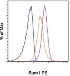
- C57Bl/6 thymocytes were stained with Anti-Mouse CD4 APC (Product # 17-0041-82) and Anti-Mouse CD8a FITC (Product # 11-0081-82), followed by intracellular staining with 0.5 µg of Rat IgG2a K Isotype Control PE (Product # 12-4321-80) (blue histogram) or 0.5 µg of Anti-Human/Mouse Runx1 PE using the Foxp3 Staining Buffer Set and protocol (Product # 00-5523-00). The histogram demonstrates Runx1 staining of CD4 SP (purple histogram) and CD8 SP (orange histogram) thymocytes.
Supportive validation
- Submitted by
- Invitrogen Antibodies (provider)
- Main image
- Experimental details
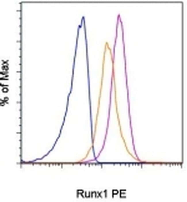
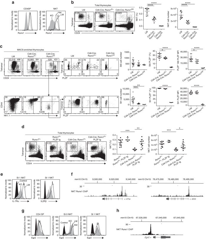
- Fig. 7 Conditional ablation of Runx1 in NKT. a Intracellular flow cytometry for Runx1 expression in thymic CD4SP and NKT cells. (LM controls (LM), solid histogram; Cd4 - Cre Runx1 fl/+ (Het), dashed histogram; Cd4 - Cre Runx1 fl/fl (KO), gray shaded .) b NKT cells in the thymus of Het and KO compared to their LM. The frequency and absolute cell number of NKT are summarized on the right panels . Data are representative of five to eight mice from four independent experiments. c Individual thymus of indicated strains were MACS-enriched using CD1d-alphaGalCer tetramers before FACS analysis of NKT developmental subsets and PLZF expression. Data are summarized from four independent experiments, with 4-12 mice in each group. d Rescue of NKT defect in Het by PLZF transgene expression. Data are representative of four to 11 mice from three independent experiments. e Representative FACS analysis of IL-7Ralpha and IL-2Rbeta expression in stage 1 NKT thymocytes from LM and KO; mean +- S.E.M. of mean fluorescence intensity (MFI) is 5535 +- 331 ( n = 4) vs 1501 +- 40 ( n = 3) for IL-7Ralpha; and 5721 +- 87 ( n = 4) vs 3040 +- 163 ( n = 3) for IL-2Rbeta, respectively. Leftmost histogram represents unstained control. f Runx1 ChIP-seq tracks at the Il7ra and Il2rb loci. g FACS analysis of Egr2 expression in CD4 SP, stage 0 and stage 1 NKT thymocytes from LM, Het and KO. Mean +- S.E.M. of Egr2 MFI: 2090 +- 84 (LM, n = 5), 1927 +- 45 (Het, n = 4) and 1506 +- 18 (KO, n = 6) in CD4 SP; 9688 +- 461
- Conjugate
- Yellow dye
- Submitted by
- Invitrogen Antibodies (provider)
- Main image
- Experimental details
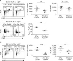
- Fig. 8 Conditional ablation of Runx1 in ILCP. a Top PLZF expression in BM Lin - IL7Ralpha + alpha4beta7 + cells from Il7ra - Cre and Il7ra - Cre Runx1 fl/fl . BM samples were MACS-enriched using anti-alpha4beta7 antibody before staining with other antibodies. Summary data of ILCP cell number and PLZF mean fluorescence intensity of ILCP cells are combined from seven independent experiments with 15 and 12 mice in each group. Bottom, Runx1 and PLZF staining in BM Lin - IL7Ralpha + alpha4beta7 + cells from Il7ra - Cre and Il7ra - Cre Runx1 fl/fl . Data are representative of three mice from each group. b PLZF expression in BM Lin - IL7Ralpha + alpha4beta7 + cells from Il7ra - Cre and Il7ra - Cre Cbfb fl/fl . Data are summarized from four independent experiments, with 8-11 mice in each group. Statistical analysis was performed using two-tailed Student's t -test. * P < 0.05, ** P < 0.01, *** P < 0.001, **** P < 0.0001
- Conjugate
- Yellow dye
