 Explore
 Validate
 Learn
Learn Western blot
Antibody data
- Antibody Data
- Antigen structure
- References [1]
- Comments [0]
- Validations
- Western blot [3]
- Immunocytochemistry [2]
Submit
Validation data
Reference
Comment
Report error
- Product number
- MA5-15814 - Provider product page

- Provider
- Invitrogen Antibodies
- Product name
- RUNX1 Monoclonal Antibody (5A1)
- Antibody type
- Monoclonal
- Antigen
- Synthetic peptide
- Description
- MA5-15814 targets RUNX1 in IF and WB applications and shows reactivity with Human samples.
- Antibody clone number
- 5A1
- Concentration
- Conc. Not Determined
Submitted references The splicing factor U2AF1 contributes to cancer progression through a noncanonical role in translation regulation.
Palangat M, Anastasakis DG, Fei DL, Lindblad KE, Bradley R, Hourigan CS, Hafner M, Larson DR
Genes & development 2019 May 1;33(9-10):482-497
Genes & development 2019 May 1;33(9-10):482-497
No comments: Submit comment
Supportive validation
- Submitted by
- Invitrogen Antibodies (provider)
- Main image
- Experimental details
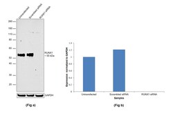
- Knockdown of RUNX1 was achieved by transfecting Jurkat with RUNX1 specific siRNAs (Silencer® select Product # s229351, s2458). Western blot analysis (Fig. a) was performed using modified whole cell extracts (1% SDS) from the RUNX1 knockdown cells (lane 3), non-specific scrambled siRNA transfected cells (lane 2) and untransfected cells (lane 1). The blot was probed with RUNX1 Monoclonal Antibody (5A1) (Product # MA5-15814, 1:1000 dilution) and Goat anti-Mouse IgG (H+L), Superclonal™ Recombinant Secondary Antibody, HRP conjugate (Product # A28177, 0.25 µg/mL, 1:4000 dilution). Densitometric analysis of this western blot is shown in histogram (Fig. b). Decrease in signal upon siRNA mediated knock down confirms that antibody is specific to RUNX1.

- Submitted by
- Invitrogen Antibodies (provider)
- Main image

- Experimental details
- Western blot was performed using Anti-RUNX1 Monoclonal Antibody (5A1), (Product # MA5-15814) and a 55 kDa band corresponding to RUNX1 along with uncharacterized bands (*) at ~70 kDa and ~200 kDa were observed in the cell lines tested. Modified whole cell extracts (1% SDS) (40 µg lysate) of Jurkat (Lane 1), HEL 92.1.7 (Lane 2), T-47D (Lane 3) and IMR32 (Lane 4) were electrophoresed using Novex® NuPAGE® 4-12 % Bis-Tris gel (Product # NP0321BOX). Resolved proteins were then transferred onto a nitrocellulose membrane (Product # IB23001) by iBlot® 2 Dry Blotting System (Product # IB21001). The blot was probed with the primary antibody (1:1000 dilution) and detected by chemiluminescence with Goat anti-Mouse IgG (H+L), Superclonal™ Recombinant Secondary Antibody, HRP (Product # A28177, 1:4000 dilution) using the iBright FL 1000 (Product # A32752). Chemiluminescent detection was performed using Novex® ECL Chemiluminescent Substrate Reagent Kit (Product # WP20005).

- Submitted by
- Invitrogen Antibodies (provider)
- Main image

- Experimental details
- Western blot analysis of RUNX1 using RUNX1 monoclonal antibody (Product # MA5-15814) in Jurkat cell lysate.

Supportive validation
- Submitted by
- Invitrogen Antibodies (provider)
- Main image

- Experimental details
- Immunofluorescence analysis of RUNX1 was performed using 70% confluent log phase Jurkat cells. The cells were fixed with 4% paraformaldehyde for 10 minutes, permeabilized with 0.1% Triton™ X-100 for 15 minutes, and blocked with 2% BSA for 1 hour at room temperature. The cells were labeled with RUNX1 Monoclonal Antibody (5A1) (Product # MA5-15814) at 1:100 dilution in 0.1% BSA, incubated at 4 degree Celsius overnight and then with Donkey anti-Mouse IgG (H+L) Highly Cross-Adsorbed Secondary Antibody, Alexa Fluor Plus 488 (Product # A32766) at a dilution of 1:2000 for 45 minutes at room temperature (Panel a: green). Nuclei (Panel b: blue) were stained with SlowFade® Gold Antifade Mountant with DAPI (Product # S36938). F-actin (Panel c: red) was stained with Rhodamine Phalloidin (Product # R415, 1:300). Panel d represents the merged image showing predominantly nuclear localization. Panel e represents control cells with no primary antibody to assess background. The images were captured at 60X magnification.

- Submitted by
- Invitrogen Antibodies (provider)
- Main image

- Experimental details
- Immunofluorescence analysis of HeLa cells using RUNX1 monoclonal antibody (Product # MA5-15814) (Green). Red: actin filaments have been labeled with phalloidin.
