Explore
 Validate
 Learn
Learn Western blot
Antibody data
- Antibody Data
- Antigen structure
- References [1]
- Comments [0]
- Validations
- Western blot [2]
- Immunocytochemistry [3]
- Immunohistochemistry [2]
- Flow cytometry [2]
Submit
Validation data
Reference
Comment
Report error
- Product number
- PA5-12409 - Provider product page

- Provider
- Invitrogen Antibodies
- Product name
- RUNX1 Polyclonal Antibody
- Antibody type
- Polyclonal
- Antigen
- Synthetic peptide
- Reactivity
- Human
- Host
- Rabbit
- Isotype
- IgG
- Vial size
- 400 μL
- Concentration
- 0.5 mg/mL
- Storage
- Store at 4°C short term. For long term storage, store at -20°C, avoiding freeze/thaw cycles.
Submitted references Recapitulation of erythropoiesis in congenital dyserythropoietic anaemia type I (CDA-I) identifies defects in differentiation and nucleolar abnormalities.
Scott C, Downes DJ, Brown JM, Beagrie R, Olijnik AA, Gosden M, Schwessinger R, Fisher CA, Rose A, Ferguson DJP, Johnson E, Hill QA, Okoli S, Renella R, Ryan K, Brand M, Hughes J, Roy NBA, Higgs DR, Babbs C, Buckle VJ
Haematologica 2021 Nov 1;106(11):2960-2970
Haematologica 2021 Nov 1;106(11):2960-2970
No comments: Submit comment
Supportive validation
- Submitted by
- Invitrogen Antibodies (provider)
- Main image

- Experimental details
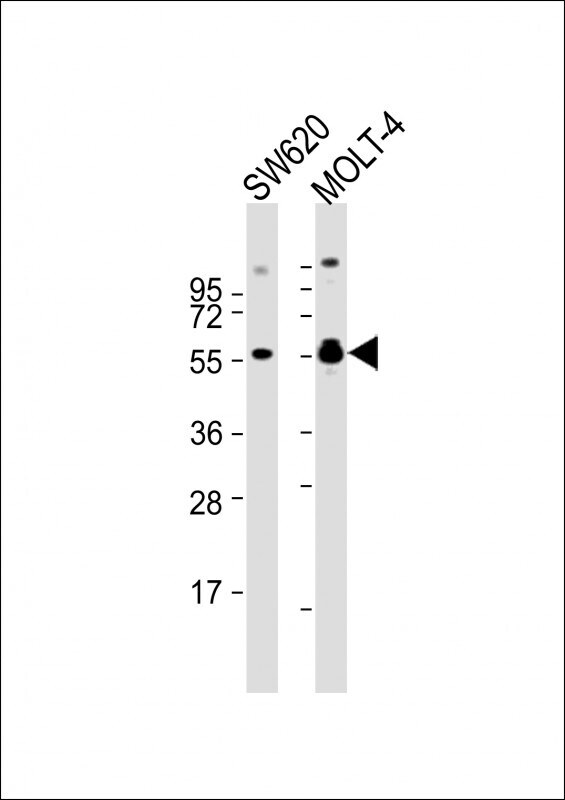
- Western blot analysis of RUNX1 in various lysates. Samples were incubated with RUNX1 polyclonal antibody (Product # PA5-12409) using a dilution of 1:2,000 followed by Goat Anti-Rabbit IgG, (H+L), Peroxidase conjugated at a dilution of 1:10,000. Lysates/proteins: 20 µg per lane. Lane 1: SW620 whole cell lysate; Lane 2: MOLT-4 whole cell lysate. Predicted band size: 49 kDa. Blocking/Dilution buffer: 5% NFDM/TBST.
- Submitted by
- Invitrogen Antibodies (provider)
- Main image

- Experimental details
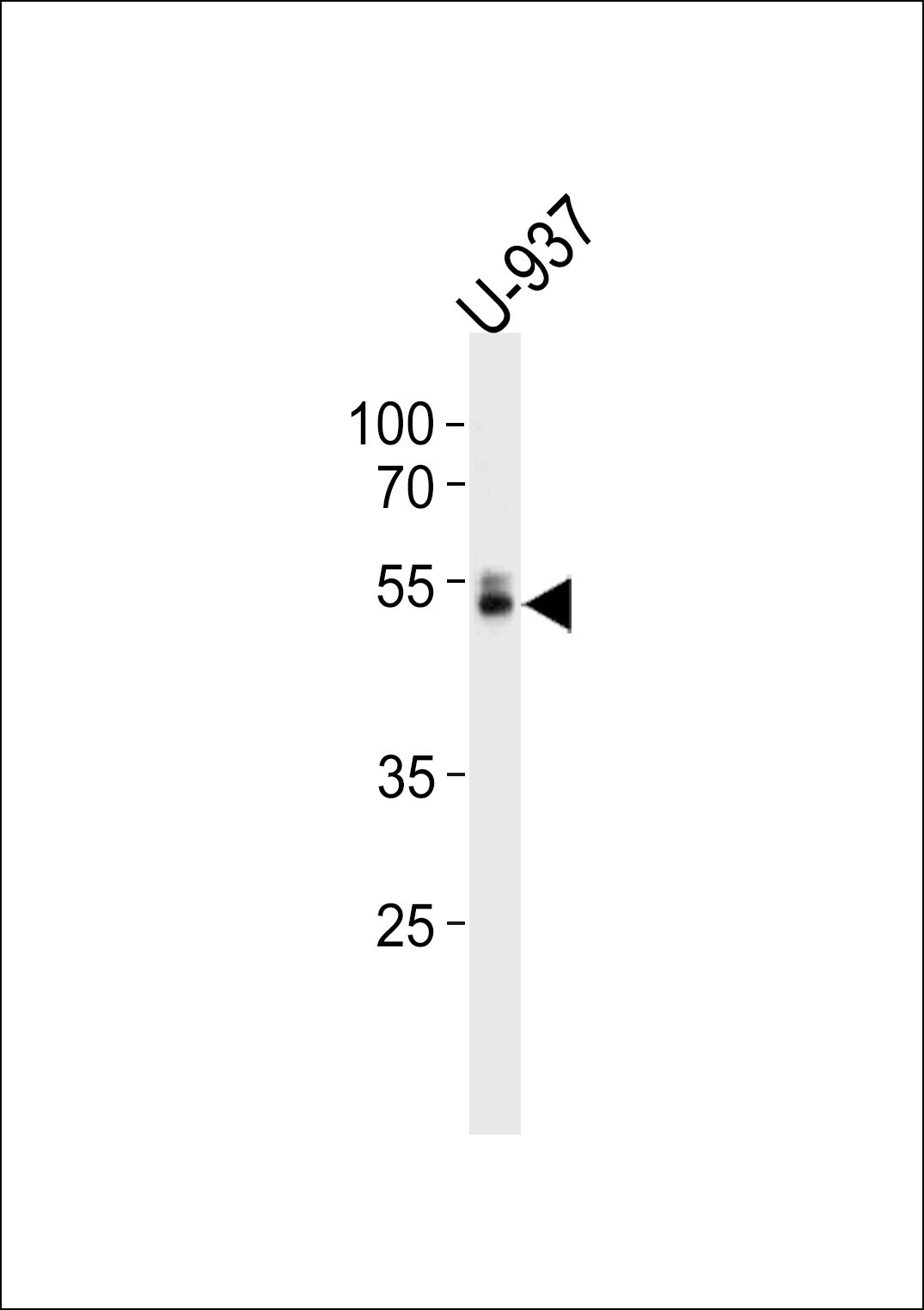
- Western blot analysis of RUNX1 in U-937 cell line lysates. Samples were incubated with RUNX1 polyclonal antibody (Product # PA5-12409). Lysates: 35 µg/lane. RUNX1 protein (arrow).
Supportive validation
- Submitted by
- Invitrogen Antibodies (provider)
- Main image
- Experimental details
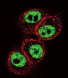
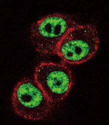
- Immunofluorescent analysis of HeLa cells using a RUNX1 polyclonal antibody (Product # PA5-12409) at a dilution of 1:10-50. Primary antibody was detected with goat anti-rabbit lgG, fluor-conjugated secondary antibody (green). Actin filaments have been labeled with red dye conjugated phalloidin.

- Submitted by
- Invitrogen Antibodies (provider)
- Main image

- Experimental details
- Immunofluorescent analysis of HeLa cells using a RUNX1 polyclonal antibody (Product # PA5-12409) at a dilution of 1:10-50. Primary antibody was detected with goat anti-rabbit lgG, fluor-conjugated secondary antibody (green). Actin filaments have been labeled with red dye conjugated phalloidin.

- Submitted by
- Invitrogen Antibodies (provider)
- Main image
- Experimental details
- Immunocytochemistry analysis of RUNX1 in HeLa cells. Samples were incubated in RUNX1 polyclonal antibody (Product # PA5-12409) followed by Alexa Fluor 488-conjugated goat anti-rabbit lgG (green). Actin filaments have been labeled with Alexa Fluor 555 phalloidin (red).

Supportive validation
- Submitted by
- Invitrogen Antibodies (provider)
- Main image
- Experimental details
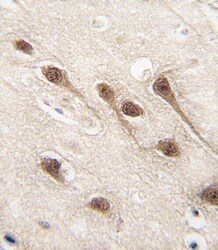
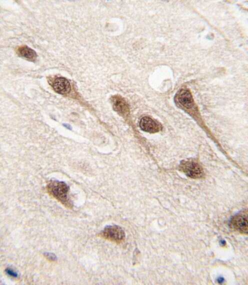
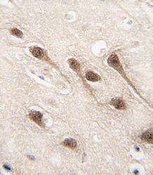
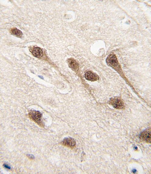
- Immunohistochemistry analysis of RUNX1 in formalin-fixed and paraffin-embedded human brain tissue. Samples were incubated with RUNX1 polyclonal antibody (Product # PA5-12409) which was peroxidase-conjugated to the secondary antibody, followed by DAB staining. This data demonstrates the use of this antibody for immunohistochemistry; clinical relevance has not been evaluated.
- Submitted by
- Invitrogen Antibodies (provider)
- Main image
- Experimental details
- Immunohistochemistry analysis of RUNX1 in formalin-fixed and paraffin-embedded human brain tissue. Samples were incubated with RUNX1 polyclonal antibody (Product # PA5-12409) which was peroxidase-conjugated to the secondary antibody, followed by DAB staining. This data demonstrates the use of this antibody for immunohistochemistry; clinical relevance has not been evaluated.
Supportive validation
- Submitted by
- Invitrogen Antibodies (provider)
- Main image
- Experimental details
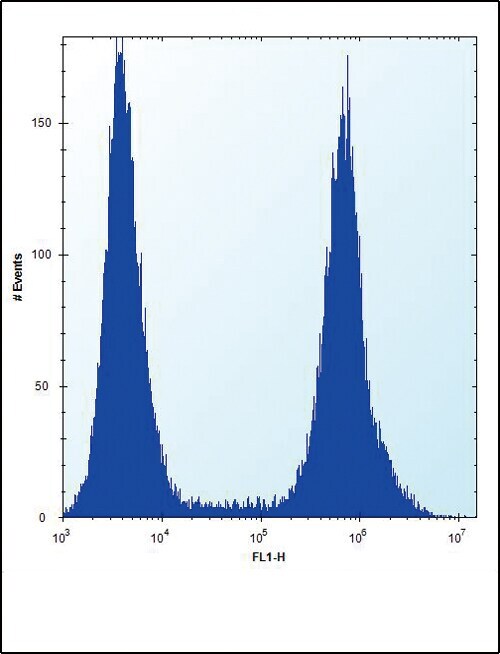
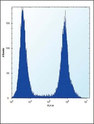
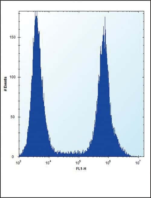
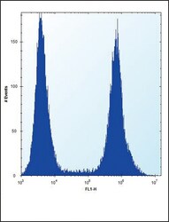
- Flow cytometry analysis of HeLa cells using a RUNX1 polyclonal antibody (Product # PA5-12409) (right) compared to a negative control cell (left) at a dilution of 1:10-50, followed by a FITC-conjugated donkey anti-rabbit antibody
- Submitted by
- Invitrogen Antibodies (provider)
- Main image
- Experimental details
- Flow cytometry of RUNX1 in Hela cells (right histogram). Samples were incubated with RUNX1 polyclonal antibody (Product # PA5-12409) followed by FITC-conjugated donkey-anti-rabbit secondary antibody. Negative control cell (left histogram).