 Explore
 Validate
 Learn
Learn Western blot
 Immunocytochemistry
Antibody data
- Antibody Data
- Antigen structure
- References [1]
- Comments [0]
- Validations
- Immunocytochemistry [5]
- Immunoprecipitation [1]
- Immunohistochemistry [1]
- Other assay [1]
Submit
Validation data
Reference
Comment
Report error
- Product number
- PA5-29597 - Provider product page

- Provider
- Invitrogen Antibodies
- Product name
- FLI1 Polyclonal Antibody
- Antibody type
- Polyclonal
- Antigen
- Recombinant full-length protein
- Description
- Recommended positive controls: Jurkat, Raji, NCI-H929, BCL-1, rat kidney. Predicted reactivity: Mouse (97%), Rat (97%), Xenopus laevis (83%), Chicken (91%), Bovine (94%). Store product as a concentrated solution. Centrifuge briefly prior to opening the vial.
- Reactivity
- Human, Rat
- Host
- Rabbit
- Isotype
- IgG
- Vial size
- 100 μL
- Concentration
- 1.22 mg/mL
- Storage
- Store at 4°C short term. For long term storage, store at -20°C, avoiding freeze/thaw cycles.
Submitted references Downregulation of Friend Leukemia Integration 1 (FLI1) follows the stepwise progression to gastric adenocarcinoma.
Del Portillo A, Komissarova EV, Bokhari A, Hills C, de Gonzalez AK, Kongkarnka S, Remotti HE, Sepulveda JL, Sepulveda AR
Oncotarget 2019 Jun 11;10(39):3852-3864
Oncotarget 2019 Jun 11;10(39):3852-3864
No comments: Submit comment
Supportive validation
- Submitted by
- Invitrogen Antibodies (provider)
- Main image
- Experimental details
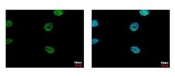
- Immunofluorescent analysis of FLI1 showing staining in the nucleus of HeLa cells. HeLa cells were fixed in 4% paraformaldehyde at RT for 15 min and stained using a FLI1 polyclonal antibody (Product # PA5-29597) diluted at 1:500. Blue: Hoechst 33343 staining.

- Submitted by
- Invitrogen Antibodies (provider)
- Main image

- Experimental details
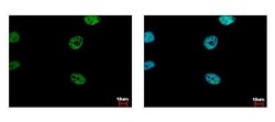
- FLI1 Polyclonal Antibody detects FLI1 protein at nucleus by immunofluorescent analysis. Sample: HeLa cells were fixed in 4% paraformaldehyde at RT for 15 min. Green: FLI1 protein stained by FLI1 Polyclonal Antibody (Product # PA5-29597) diluted at 1:500. Blue: Hoechst 33343 staining.

- Submitted by
- Invitrogen Antibodies (provider)
- Main image

- Experimental details
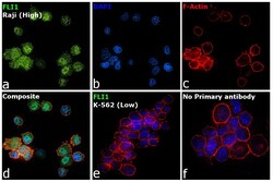
- Immunofluorescence analysis of Friend leukemia integration 1 transcription factor was performed using 70% confluent log phase Raji cells. The cells were fixed with 4% paraformaldehyde for 10 minutes, permeabilized with 0.1% Triton™ X-100 for 15 minutes, and blocked with 2% BSA for 45 minutes at room temperature. The cells were labeled with FLI1 Polyclonal Antibody (Product # PA5-29597) at (1:100 dilution) in 0.1% BSA, incubated at 4-degree Celsius overnight, and labeled with Donkey anti-Rabbit IgG (H+L) Highly Cross-Adsorbed Secondary Antibody, Alexa Fluor Plus 488 (Product # A32790), (1:2000 dilution), for 45 minutes at room temperature (Panel a: Green). Nuclei (Panel b: Blue) were stained with ProLong™ Diamond Antifade Mountant with DAPI (Product # P36962). F-actin (Panel c: Red) was stained with Rhodamine Phalloidin (Product # R415, 1:300). Panel d represents the merged image showing nuclear localization. Panel e represents K-562 cells with no expression of FLI1. Panel f represents control cells with no primary antibody to assess the background. The images were captured at 60X magnification.

- Submitted by
- Invitrogen Antibodies (provider)
- Main image
- Experimental details
- FLI1 Polyclonal Antibody detects FLI1 protein at nucleus by immunofluorescent analysis. Sample: HeLa cells were fixed in 4% paraformaldehyde at RT for 15 min. Green: FLI1 protein stained by FLI1 Polyclonal Antibody (Product # PA5-29597) diluted at 1:500. Blue: Hoechst 33343 staining.

Supportive validation
- Submitted by
- Invitrogen Antibodies (provider)
- Main image

- Experimental details
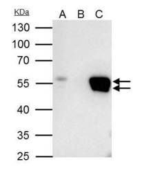
- FLI1 Polyclonal Antibody immunoprecipitates FLI1 protein in IP experiments. IP samples: Raji whole cell extract. A. 40 µg Raji whole cell extract. B. Control with 4 µg of preimmune Rabbit IgG. C. Immunoprecipitation of FLI1 protein by 4 µg FLI1 Polyclonal Antibody (Product # PA5-29597). 10 % SDS-PAGE. The immunoprecipitated FLI1 protein was detected by FLI1 Polyclonal Antibody (Product # PA5-29597) diluted at 1:500.

Supportive validation
- Submitted by
- Invitrogen Antibodies (provider)
- Main image
- Experimental details
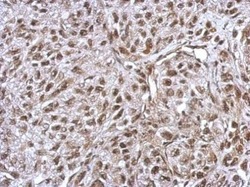
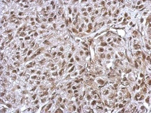
- Immunohistochemical analysis of paraffin-embedded SkHep1 xenograft, using FLI1 (Product # PA5-29597) antibody at 1:500 dilution. Antigen Retrieval: EDTA based buffer, pH 8.0, 15 min.
Supportive validation
- Submitted by
- Invitrogen Antibodies (provider)
- Main image
- Experimental details
- FLI1 Polyclonal Antibody immunoprecipitates FLI1 protein in IP experiments. IP samples: Raji whole cell extract. A. 40 µg Raji whole cell extract. B. Control with 4 µg of preimmune Rabbit IgG. C. Immunoprecipitation of FLI1 protein by 4 µg FLI1 Polyclonal Antibody (Product # PA5-29597). 10 % SDS-PAGE. The immunoprecipitated FLI1 protein was detected by FLI1 Polyclonal Antibody (Product # PA5-29597) diluted at 1:500.
