 Explore
 Validate
 Learn
Learn Western blot
 ELISA
ELISAAntibody data
- Antibody Data
- Antigen structure
- References [1]
- Comments [0]
- Validations
- Western blot [2]
- Immunocytochemistry [1]
- Immunohistochemistry [1]
- Flow cytometry [2]
- Other assay [2]
Submit
Validation data
Reference
Comment
Report error
- Product number
- MA1-19319 - Provider product page

- Provider
- Invitrogen Antibodies
- Product name
- Vimentin Monoclonal Antibody (VI-RE/1)
- Antibody type
- Monoclonal
- Antigen
- Other
- Description
- This antibody reacts with human vimentin, a 57 kDa intermediate filament intracellular protein expressed on a wide variety of mesenchymal and mesodermal cell types. This antibody will not cross-react with porcine or mouse.
- Reactivity
- Human
- Host
- Mouse
- Isotype
- IgG
- Antibody clone number
- VI-RE/1
- Vial size
- 100 µg
- Concentration
- 1 mg/mL
- Storage
- 4° C, do not freeze
Submitted references Carcinoma cells that have undergone an epithelial-mesenchymal transition differentiate into endothelial cells and contribute to tumor growth.
Sphyris N, King C, Hoar J, Werden SJ, Vijay GV, Miura N, Gaharwar A, Sarkar TR
Oncotarget 2021 Apr 13;12(8):823-844
Oncotarget 2021 Apr 13;12(8):823-844
No comments: Submit comment
Supportive validation
- Submitted by
- Invitrogen Antibodies (provider)
- Main image
- Experimental details
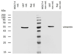
- Western blotting analysis of human vimentin using mouse monoclonal antibody VI-RE/1 on lysates of MOLT-4 cell line (low expression), U87 cell line (positive) and Raji cell line (negative control) under non-reducing and reducing conditions. Nitrocellulose membrane was probed with 2µg/mL of mouse anti-vimentin Monoclonal antibody (Product # MA1-19319) followed by IRDye800-conjugated anti-mouse secondary antibody. A specific band was detected for vimentin at approximately 55kDa.

- Submitted by
- Invitrogen Antibodies (provider)
- Main image

- Experimental details
- Western blot analysis was performed on Whole cell extracts (30 µg lysate) of HeLa (Lane 1), A-431 (Lane 2), SHSY-5Y (Lane 3), Hep G2 (Lane 4) and NIH/3T3 (Lane 5). The blot was probed with Anti-Vimentin Monoclonal Antibody (Product # MA1-19319, 1:2000 dilution) and detected by chemiluminescence using Goat anti-Mouse IgG (H+L) Superclonal™ Secondary Antibody, HRP conjugate (Product # A28177, 0.25 µg/ml, 1:4000 dilution). A 54 kDa band corresponding to Vimentin was observed across all the human cell lines positive for Vimentin (Lanes 1 and 3) but not mouse cell line (Lane 5), while this band was absent in the cell lines which do not express Vimentin protein (Lanes 2 and 4).

Supportive validation
- Submitted by
- Invitrogen Antibodies (provider)
- Main image
- Experimental details
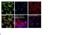
- Immunofluorescence analysis of Vimentin was performed using 70% confluent log phase HeLa cells. The cells were fixed with 4% paraformaldehyde for 10 minutes, permeabilized with 0.1% Triton™ X-100 for 10 minutes, and blocked with 1% BSA for 1 hour at room temperature. The cells were labeled with Vimentin Mouse Monoclonal Antibody (Product # MA1-19319) at 5 µg/mL in 0.1% BSA and incubated overnight at 4 degree Celsius and then labeled with Goat anti-Mouse IgG (H+L) Superclonal™ Secondary Antibody, Alexa Fluor® 488 conjugate (Product # A28175) at a dilution of 1:2000 for 45 minutes at room temperature (Panel a: green). Nuclei (Panel b: blue) were stained with SlowFade® Gold Antifade Mountant with DAPI (Product # S36938). F-actin (Panel c: red) was stained with Rhodamine Phalloidin (Product # R415, 1:300). Panel d represents the merged image showing cytoplasmic, cytoskeletal and nuclear localization. Panel e represents negative control, A-431 cells. Panel f represents control cells with no primary antibody to assess background. The images were captured at 60X magnification.

Supportive validation
- Submitted by
- Invitrogen Antibodies (provider)
- Main image

- Experimental details
- Immunohistochemistry staining (paraffin sections) of vimentin in human liver using mouse Monoclonal antibody VI-RE/1 (Product # MA1-19319) using a dilution of 1:400, detected with GAM IgG-Alexa Fluor®488 (diluted 1:200; green), cell nuclei stained with PI (1 µg/mL; orange).

Supportive validation
- Submitted by
- Invitrogen Antibodies (provider)
- Main image

- Experimental details
- Intracellular flow cytometry analysis of Vimentin expression in LEP-19 human fibroblast cell line using anti-human Vimentin monoclonal antibody (Product # MA1-19319). Overlay with Isotype mouse IgG1 control

- Submitted by
- Invitrogen Antibodies (provider)
- Main image
- Experimental details
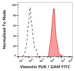
- Flow cytometry analysis (intracellular staining) of vimentin expression in ESS-1 cells using anti-human vimentin (VI-RE/1) purified Monoclonal antibody (Product # MA1-19319), GAM-FITC. Negative control: human lymphocytes.

Supportive validation
- Submitted by
- Invitrogen Antibodies (provider)
- Main image

- Experimental details
- Figure 2 MCF-7 tumor outgrowth is accompanied by increased expression of markers of EMT and hypoxia. RFP/luciferase-labeled MCF-7 cells were orthotopically injected into recipient NOD/SCID hosts, and tumors were allowed to grow to until their size reached approximately

- Submitted by
- Invitrogen Antibodies (provider)
- Main image

- Experimental details
- Figure 4 HMLE-Snail cells gain CD31 expression and promote acquisition of mesenchymal traits by admixed MCF-7 cells. RFP/luciferase-labeled MCF-7 cells (0.5 x 10 6 ) were co-mixed with 0.5 x 10 6 MCF-7, HMLE-vector, or HMLE-Snail cells, and orthotopically implanted into female NOD/SCID mice. Ten weeks post implantation, the tumors were harvested and processed for immunofluorescent staining. n = 5 mice per group. ( A , B ) Tumor core sections were co-stained with antibodies directed against RFP (red) and either E-cadherin (A) or vimentin (B), both pseudo-colored green. Nuclei were counterstained with DAPI (blue). Right panels are merged images of individual channels. Note the progressive reduction of E-cadherin expression, commensurate with the attenuation of the honeycomb-like membrane staining pattern across these tumor cores and the inversely-correlated augmented vimentin staining. ( C ) Sections from the tumor cores were co-stained with antibodies directed against the SV40 large-T antigen (green) and human CD31 (red). Nuclei were counterstained with DAPI (blue). Right panels are merged images of individual channels. Arrows indicate SV40 large-T antigen/CD31 double-positive cells lining the lumens of vascular structures in tumors formed by admixed MCF-7/HMLE-Snail cells. Boxed areas represent high-magnification images of selected encircled areas. Scale bars, 100 mum. Representative images are shown.
