 Explore
 Validate
 Learn
Learn Western blot
 Flow cytometry
Antibody data
- Antibody Data
- Antigen structure
- References [6]
- Comments [0]
- Validations
- Western blot [3]
- Immunocytochemistry [1]
- Immunohistochemistry [8]
- Other assay [3]
Submit
Validation data
Reference
Comment
Report error
- Product number
- OMA1-06001 - Provider product page

- Provider
- Invitrogen Antibodies
- Product name
- Vimentin Monoclonal Antibody (RV202)
- Antibody type
- Monoclonal
- Antigen
- Other
- Description
- OMA1-06001 detects vimentin from mouse, rat, hamster, chicken, goat, canine, monkey, zebrafish, porcine and human tissues and cells. OMA1-06001 has been successfully used in Western blot, immunofluorescence, immunocytochemistry, flow cytometry, and immunohistochemistry (frozen) procedures. By Western blot, this antibody detects a ~57 kDa protein representing vimentin from human mesenchymal cells. OMA1-06001 antigen is cytoskeletal vimentin extract of calf lens. Store at 4ºC or at -20ºC if preferred. Avoid freeze-thaw cycles.
- Reactivity
- Human, Mouse, Rat, Bovine, Canine, Chicken/Avian, Goat, Hamster, Porcine, Xenopus, Zebrafish
- Host
- Mouse
- Isotype
- IgG
- Antibody clone number
- RV202
- Vial size
- 100 µL
- Concentration
- 1 mg/mL
- Storage
- Store at 4°C short term. For long term storage, store at -20°C, avoiding freeze/thaw cycles.
Submitted references Renalase and its receptor, PMCA4b, are expressed in the placenta throughout the human gestation.
Effect of canonical NF-κB signaling pathway on the differentiation of rat dental epithelial stem cells.
A systems-approach reveals human nestin is an endothelial-enriched, angiogenesis-independent intermediate filament protein.
Schwann cells secrete extracellular vesicles to promote and maintain the proliferation and multipotency of hDPCs.
Disruption of kif3a results in defective osteoblastic differentiation in dental mesenchymal stem/precursor cells via the Wnt signaling pathway.
Osteogenic differentiation and gene expression profile of human dental follicle cells induced by human dental pulp cells.
Wang M, Silva T, Toothaker JM, McCourt BT, Shugrue C, Desir G, Gorelick F, Konnikova L
Scientific reports 2022 Mar 23;12(1):4953
Scientific reports 2022 Mar 23;12(1):4953
Effect of canonical NF-κB signaling pathway on the differentiation of rat dental epithelial stem cells.
Liang Y, Chen G, Yang Y, Li Z, Chen T, Sun W, Yu M, Pan K, Guo W, Tian W
Stem cell research & therapy 2019 May 20;10(1):139
Stem cell research & therapy 2019 May 20;10(1):139
A systems-approach reveals human nestin is an endothelial-enriched, angiogenesis-independent intermediate filament protein.
Dusart P, Fagerberg L, Perisic L, Civelek M, Struck E, Hedin U, Uhlén M, Trégouët DA, Renné T, Odeberg J, Butler LM
Scientific reports 2018 Oct 2;8(1):14668
Scientific reports 2018 Oct 2;8(1):14668
Schwann cells secrete extracellular vesicles to promote and maintain the proliferation and multipotency of hDPCs.
Li Z, Liang Y, Pan K, Li H, Yu M, Guo W, Chen G, Tian W
Cell proliferation 2017 Aug;50(4)
Cell proliferation 2017 Aug;50(4)
Disruption of kif3a results in defective osteoblastic differentiation in dental mesenchymal stem/precursor cells via the Wnt signaling pathway.
Jiang S, Chen G, Feng L, Jiang Z, Yu M, Bao J, Tian W
Molecular medicine reports 2016 Sep;14(3):1891-900
Molecular medicine reports 2016 Sep;14(3):1891-900
Osteogenic differentiation and gene expression profile of human dental follicle cells induced by human dental pulp cells.
Park SJ, Bae HS, Park JC
Journal of molecular histology 2015 Feb;46(1):93-106
Journal of molecular histology 2015 Feb;46(1):93-106
No comments: Submit comment
Supportive validation
- Submitted by
- Invitrogen Antibodies (provider)
- Main image

- Experimental details
- Western blot was performed using Anti-Vimentin Monoclonal Antibody (RV202) (Product # OMA1-06001) and a ~58kDa band corresponding to vimentin was observed across cell lines tested except in A-431. Whole cell extracts (30 µg lysate) of HeLa (Lane 1), SH-SY5Y (Lane 2), A549 (Lane 3), A-431 (Lane 4) were electrophoresed using NuPAGE™ 4-12% Bis-Tris Protein Gel (Product # NP0322BOX), 12 well. Resolved proteins were then transferred onto a nitrocellulose membrane (Product # IB23001) by iBlot® 2 Dry Blotting System (Product # IB21001). The blot was probed with the primary antibody (1:1000) and detected by chemiluminescence with Goat anti-Mouse IgG (H+L) Superclonal™ Recombinant Secondary Antibody, HRP (Product # A28177,1:20000) using the iBright™ FL1500 Imaging System (Product # A44115). Chemiluminescent detection was performed using SuperSignal™ West Pico PLUS Chemiluminescent Substrate (Product # 34580).

- Submitted by
- Invitrogen Antibodies (provider)
- Main image

- Experimental details
- CRISPR-Cas9 mediated genome editing ofVimentin (as confirmed by next generation sequencing) was achieved by using LentiArray™ Lentiviral sgRNA (Product # A32042, Assay ID CRISPR931291_LV) and LentiArray Cas9 Lentivirus (Product # A32064). Fig (a) Western blot analysis of Vimentin was performed by loading 30 µg of HeLa Wild Type (Lane 1), HeLa Cas9 (Lane 2) and HeLa Cas9 cells transduced with Vimentin Lentiviral sgRNA (Lane 3) whole cell extracts. The samples were electrophoresed using NuPAGE™ Novex™ 4-12% Bis-Tris Protein Gel (Product # NP0322BOX). Resolved proteins were then transferred onto a nitrocellulose membrane (Product # IB23001) by iBlot® 2 Dry Blotting System (Product # IB21001). The blot was probed with Anti-Vimentin Monoclonal Antibody (RV202) (Product # OMA1-06001) using 1:1,000 dilution and Goat anti-Mouse IgG (H+L) Superclonal™ Recombinant Secondary Antibody, HRP (Product # A28177 1:4,000 dilution). Chemiluminescent detection was performed using Novex® ECL Chemiluminescent Substrate Reagent Kit (Product # WP20005).Even though NGS analysis determine the clone as partial KO, there was complete loss of signal in sgRNA transduced cells using the LentiArray™ CRISPR product line confirming that the antibody is specific to Vimentin (Fig (b)).

- Submitted by
- Invitrogen Antibodies (provider)
- Main image

- Experimental details
- Western blot was performed using Anti-Vimentin Polyclonal Antibody (Product # OMA1-06001) and a 53 kDa band corresponding to Vimentin was observed across all the cell lines tested except in MCF7 and T-47D. Whole cell extracts (30 µg lysate) of MDA-MB-231 (Lane 1), MCF-7 (Lane 2), T-47D (Lane 3), A549 (Lane 4), HeLa (Lane 5), SH-SY5Y (Lane 6) and A-431 (Lane 7) were electrophoresed using NuPAGE™ 4-12% Bis-Tris Protein Gel (Product # NP0322BOX). Resolved proteins were then transferred onto a nitrocellulose membrane (Product # IB23001) by iBlot® 2 Dry Blotting System (Product # IB21001). The blot was probed with the primary antibody (1:1000 dilution) and detected by chemiluminescence with Goat anti-Mouse IgG (H+L) Superclonal™ Secondary Antibody, HRP (Product # A28177, 1:4000 dilution) using the iBright FL 1000 (Product # A32752). Chemiluminescent detection was performed using Novex® ECL Chemiluminescent Substrate Reagent Kit (Product # WP20005).

Supportive validation
- Submitted by
- Invitrogen Antibodies (provider)
- Main image

- Experimental details
- Immunofluorescence analysis of Vimentin was performed using 70% confluent log phase MDA-MB-231 cells. The cells were fixed with 4% paraformaldehyde for 10 minutes, permeabilized with 0.1% Triton™ X-100 for 10 minutes, and blocked with 2% BSA for 10 minutes at room temperature. The cells were labeled with Vimentin Polyclonal Antibody (Product # OMA1-06001) at 1:100 dilution in 0.1% BSA, incubated at 4 degree celsius overnight and then labeled with Donkey anti-Mouse IgG (H+L) Highly Cross-Adsorbed Secondary Antibody, Alexa Fluor Plus 488 (Product # A32766), (1:2000 dilution), for 45 minutes at room temperature (Panel a: Green). Nuclei (Panel b: blue) were stained with SlowFade® Gold Antifade Mountant with DAPI (Product # S36938). F-actin (Panel c: Red) was stained with Rhodamine Phalloidin (Product # R415, 1:300). Panel d represents the merged image showing cytoskeletal localization. Panel e represents MCF7 cells having no expression of Vimentin. Panel f represents control cells with no primary antibody to assess background. The images were captured at 60X magnification.

Supportive validation
- Submitted by
- Invitrogen Antibodies (provider)
- Main image

- Experimental details
- Immunofluorescent analysis of 1 month old zebrafish embryo using Vimentin monoclonal antibody (Product # OMA1-06001).

- Submitted by
- Invitrogen Antibodies (provider)
- Main image
- Experimental details
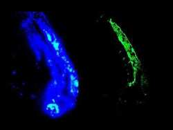
- Immunofluorescent analysis of developing neural tube in 2 days old zebrafish embryo. using Vimentin monoclonal antibody (Product # OMA1-06001). Left panel: DAPI-staining of cell nuclei, providing an overview of the tissue section used for immunostaining

- Submitted by
- Invitrogen Antibodies (provider)
- Main image

- Experimental details
- Immunofluorescent analysis of 1 month old zebrafish embryo using Vimentin monoclonal antibody (Product # OMA1-06001).

- Submitted by
- Invitrogen Antibodies (provider)
- Main image

- Experimental details
- Immunohistochemical staining of frozen section of swine colon using Vimentin monoclonal antibody (Product # OMA1-06001) (1:200).
- Submitted by
- Invitrogen Antibodies (provider)
- Main image

- Experimental details
- Immunohistochemical staining of frozen section of swine colon immunostained with Vimentin monoclonal antibody (Product # OMA1-06001) (1:200).
- Submitted by
- Invitrogen Antibodies (provider)
- Main image

- Experimental details
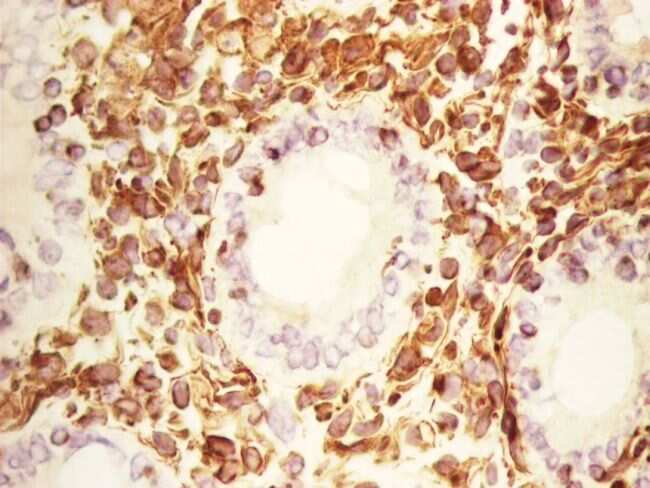
- Immunohistochemistry on paraffin section of human colon stained with Vimentin monoclonal antibody (Product # OMA1-06001).

- Submitted by
- Invitrogen Antibodies (provider)
- Main image

- Experimental details
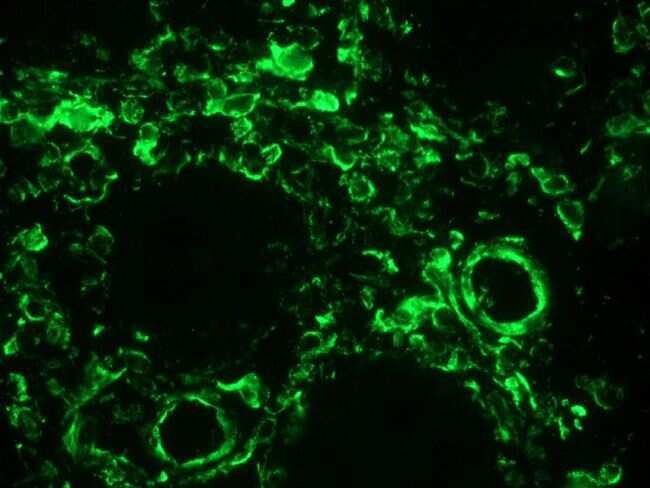
- Immunohistochemistry of frozen section of swine colon showing positive staining in connective tissue cells and no reactivity in epithelial cells. Primary antibody was a Vimentin monoclonal antibody (Product # OMA1-06001).

- Submitted by
- Invitrogen Antibodies (provider)
- Main image

- Experimental details
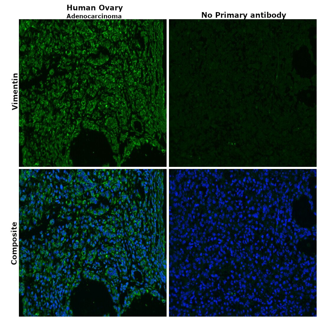
- Immunohistochemical analysis of vimentin was performed using formalin-fixed paraffin-embedded human ovary adenocarcinoma tissue sections. To expose the target protein, heat-induced epitope retrieval was performed on de-paraffinized sections using eBioscience™ IHC Antigen Retrieval Solution - Low pH (10X) (Product # 00-4955-58) diluted to 1X solution in water in a decloaking chamber at 110 degree celsius for 15 minutes. Following antigen retrieval, the sections were blocked with 2% normal goat serum in 1X PBS for 45 minutes at room temperature and then probed with or without Vimentin Monoclonal Antibody (RV202) (Product # OMA1-06001) at 1:100 dilution in 0.1% normal goat serum overnight at 4 degree celsius in a humidified chamber. Detection was performed using Goat anti-Mouse IgG (H+L) Highly Cross-Adsorbed Secondary Antibody, Alexa Fluor Plus 488 (Product # A32723) at a dilution of 1:2000 in 0.1% normal goat serum for 45 minutes at room temperature. Nuclei were stained with DAPI (Product # D1306) and the sections were mounted using ProLong™ Glass Antifade Mountant (Product # P36984). The images were captured on EVOS™ M7000 Imaging System (Product # AMF7000) at 20X magnification.
Supportive validation
- Submitted by
- Invitrogen Antibodies (provider)
- Main image

- Experimental details
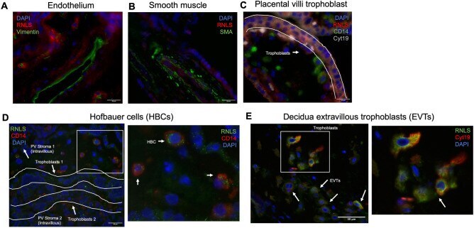
- Figure 4 RNLS immunoreactivity is present in trophoblasts and macrophages within placental villi and decidua. (A) Immunofluorescence co-labeling with RNLS (red), vimentin (green, endothelium), DAPI (blue) of placental villi at 60x. (B) RNLS (red), SMA (green, smooth muscle actin, smooth muscle), and DAPI (blue) of placental villi at 60x. (C) RNLS (red), CD14 (green, Hofbauer cell), Cyt19 (white, cytokeratin 19, trophoblast), DAPI (blue) in placental villi at 60x. (D) RNLS (green), CD14 (red, Hofbauer cell) and DAPI (blue) of placental villi trophoblasts at 60x. (E) RNLS (green), Cyt19 (red, extravillous trophoblast, EVT), DAPI (blue) of placental decidua at 60x.
- Submitted by
- Invitrogen Antibodies (provider)
- Main image

- Experimental details
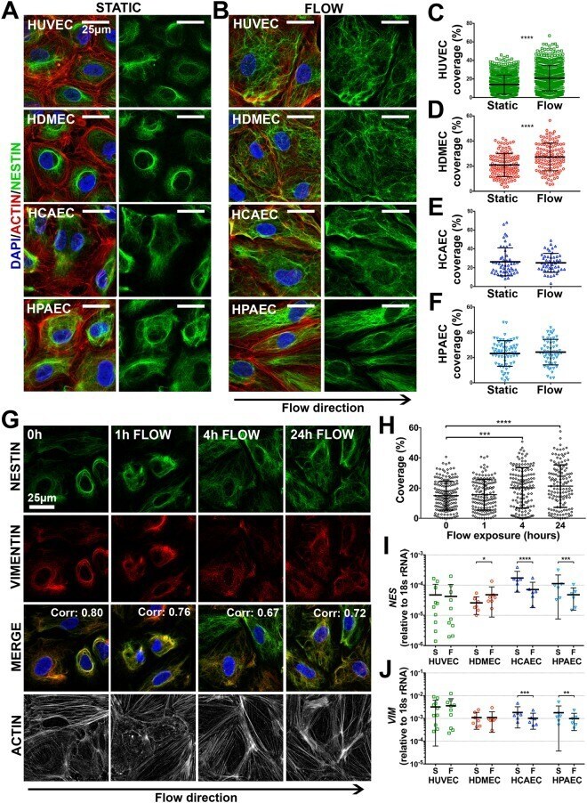
- Figure 4 EC nestin is regulated by laminar shear stress in vitro . Immunofluorescence staining of nestin in HUVEC, HDMEC, HCAEC and HPAEC cultured under ( A ) static conditions, or ( B ) 10 dyne/cm 2 laminar shear stress for 24 hours. Quantification of nestin spatial distribution in ( C ) HUVEC, ( D ) HDMEC, ( E ) HCAEC and ( F ) HPAEC. Each point represents an individual cell (n = 3-19 different experiments) Unpaired t-test *p-value < 0.05, **
- Submitted by
- Invitrogen Antibodies (provider)
- Main image

- Experimental details
- Figure 1 Evaluation of the biological characteristics of hDFCs and hDPCs. The two types of cell were positive for vimentin and Stro-1, but negative for the epithelial marker, CK-14. Scale bar=20 u m. hDFCs, human dental follicle cells; hDPCs, human dental pulp cells.
