 Explore
 Validate
 Learn
Learn Western blot
 Immunohistochemistry
Antibody data
- Antibody Data
- Antigen structure
- References [2]
- Comments [0]
- Validations
- Western blot [1]
- Immunocytochemistry [1]
Submit
Validation data
Reference
Comment
Report error
- Product number
- ABIN1580467 - Provider product page

- Provider
- antibodies-online
- Product name
- anti-Vimentin (VIM) antibody
- Antibody type
- Monoclonal
- Antigen
- Other
- Description
- affinity purified antibody
- Reactivity
- Human, Mouse, Rat, Bovine, Porcine
- Host
- Mouse
- Isotype
- IgG
- Antibody clone number
- 2A52
- Vial size
- 100 μL
- Concentration
- 1 mg/mL
- Storage
- Store at 4°C short term or -20°C long term.
- Handling
- Avoid repeated freezing and thawing.
Submitted references Dominant cataract formation in association with a vimentin assembly disrupting mutation.
Different intermediate-sized filaments distinguished by immunofluorescence microscopy.
Müller M, Bhattacharya SS, Moore T, Prescott Q, Wedig T, Herrmann H, Magin TM
Human molecular genetics 2009 Mar 15;18(6):1052-7
Human molecular genetics 2009 Mar 15;18(6):1052-7
Different intermediate-sized filaments distinguished by immunofluorescence microscopy.
Franke WW, Schmid E, Osborn M, Weber K
Proceedings of the National Academy of Sciences of the United States of America 1978 Oct;75(10):5034-8
Proceedings of the National Academy of Sciences of the United States of America 1978 Oct;75(10):5034-8
No comments: Submit comment
Supportive validation
- Submitted by
- antibodies-online (provider)
- Main image
- Experimental details
- Western blot of crude extract of the human carcinoma HeLa cell line. Lane 18 was probed with MCA-2D1 antibody. Note the strong clean band at the expected molecular weight of 50 kDa. Lane 17 was probed with another monoclonal antibody to vimentin, our ABIN1580467 clone. Lane 15 was probed with our antibody to actin, giving an SDS-PAGE molecular weight of 42 kDa, and lane 16 with MCA-3G12, our antibody to 14-3-3? (14-3-3 eta), which has an SDS-PAGE molecular weight of 28 kDa.

Supportive validation
- Submitted by
- antibodies-online (provider)
- Main image

- Experimental details
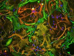
- View of mixed neuron/glial cultures stained with CPCA-Vim (green) and ?s rabbit antibody to GFAP antibody (RPCA-GFAP, red). Vimentin is expressed alone in fibroblastic and endothelial cells, which are the flattened cells in the middle of the image which appear green. Astrocytes may express primarily GFAP, or GFAP and vimentin, and so appear red (GFAP only) or golden yellow (GFAP and Vimentin). In cells which express both GFAP and vimentin, the two protein assemble to produce heteropolymer filaments.
