Explore
 Validate
 Learn
Learn Western blot
 Immunocytochemistry
Antibody data
- Antibody Data
- Antigen structure
- References [46]
- Comments [0]
- Validations
- Western blot [24]
- Immunocytochemistry [6]
- Immunoprecipitation [1]
- Immunohistochemistry [10]
Submit
Validation data
Reference
Comment
Report error
- Product number
- GTX100619 - Provider product page

- Provider
- GeneTex
- Proper citation
- GeneTex Cat#GTX100619, RRID:AB_1952557
- Product name
- Vimentin antibody
- Antibody type
- Polyclonal
- Reactivity
- Human, Mouse, Rat
- Host
- Rabbit
Submitted references Sympathetic innervation contributes to perineural invasion of salivary adenoid cystic carcinoma via the β2-adrenergic receptor.
Methyl-CpG Binding Domain Protein 2 Inhibits the Malignant Characteristic of Lung Adenocarcinoma through the Epigenetic Modulation of 10 to 11 Translocation 1 and miR-200s.
Fucoidan Inhibits the Proliferation of Leiomyoma Cells and Decreases Extracellular Matrix-Associated Protein Expression.
PEGylated liposome-encapsulated rhenium-188 radiopharmaceutical inhibits proliferation and epithelial-mesenchymal transition of human head and neck cancer cells in vivo with repeated therapy.
UHRF1 regulates CDH1 via promoter associated non-coding RNAs in prostate cancer cells.
Periostin blockade overcomes chemoresistance via restricting the expansion of mesenchymal tumor subpopulations in breast cancer.
Daily therapy with a slow-releasing H2S donor GYY4137 enables early functional recovery and ameliorates renal injury associated with urinary obstruction.
Transforming growth factor alpha promotes tumorigenesis and regulates epithelial-mesenchymal transition modulation in colon cancer.
DNA methylation variations are required for epithelial-to-mesenchymal transition induced by cancer-associated fibroblasts in prostate cancer cells.
Role of estrogen receptors and Src signaling in mechanisms of bone metastasis by estrogen receptor positive breast cancers.
SB-T-121205, a next-generation taxane, enhances apoptosis and inhibits migration/invasion in MCF-7/PTX cells.
Sulfiredoxin may promote metastasis and invasion of cervical squamous cell carcinoma by epithelial-mesenchymal transition.
VLDL and LDL, but not HDL, promote breast cancer cell proliferation, metastasis and angiogenesis.
CYT-Rx20 inhibits ovarian cancer cells in vitro and in vivo through oxidative stress-induced DNA damage and cell apoptosis.
3'UTR polymorphisms of carbonic anhydrase IX determine the miR-34a targeting efficiency and prognosis of hepatocellular carcinoma.
Elevation of YAP promotes the epithelial-mesenchymal transition and tumor aggressiveness in colorectal cancer.
The inhibition of lung cancer cell migration by AhR-regulated autophagy.
MicroRNA-613 suppresses proliferation, migration and invasion of osteosarcoma by targeting c-MET.
Loss of the SWI/SNF ATPase subunits BRM and BRG1 drives lung cancer development.
Extracellular Visfatin-Promoted Malignant Behavior in Breast Cancer Is Mediated Through c-Abl and STAT3 Activation.
Oxystressed tumor microenvironment potentiates epithelial to mesenchymal transition and alters cellular bioenergetics towards cancer progression.
Suberoylanilide hydroxamic acid represses glioma stem-like cells.
SARS coronavirus papain-like protease induces Egr-1-dependent up-regulation of TGF-β1 via ROS/p38 MAPK/STAT3 pathway.
Aberrant DNA hypomethylation of miR-196b contributes to migration and invasion of oral cancer.
PFA fixation enables artifact-free super-resolution imaging of the actin cytoskeleton and associated proteins.
MiR-206 suppresses epithelial mesenchymal transition by targeting TGF-β signaling in estrogen receptor positive breast cancer cells.
Inhibition of colon cancer cell growth by nanoemulsion carrying gold nanoparticles and lycopene.
Long non-coding RNA AOC4P suppresses hepatocellular carcinoma metastasis by enhancing vimentin degradation and inhibiting epithelial-mesenchymal transition.
Proteomic analysis reveals novel common genes modulated in both replicative and stress-induced senescence.
PGRMC1 contributes to doxorubicin-induced chemoresistance in MES-SA uterine sarcoma.
MEF2B mutations in non-Hodgkin lymphoma dysregulate cell migration by decreasing MEF2B target gene activation.
Antroquinonol from Antrodia Camphorata suppresses breast tumor migration/invasion through inhibiting ERK-AP-1- and AKT-NF-κB-dependent MMP-9 and epithelial-mesenchymal transition expressions.
Pigment Epithelial-Derived Factor Peptide Facilitates the Regeneration of a Functional Limbus in Rabbit Partial Limbal Deficiency.
Notch2 controls prolactin and insulin-like growth factor binding protein-1 expression in decidualizing human stromal cells of early pregnancy.
FOXF1 mediates mesenchymal stem cell fusion-induced reprogramming of lung cancer cells.
Epithelial-mesenchymal transition during invasion of cutaneous squamous cell carcinoma is paralleled by AKT activation.
Notch signaling plays a critical role in motility and differentiation of human first-trimester cytotrophoblasts.
Mesenchymal stem cell characteristics of dental pulp and periodontal ligament stem cells after in vivo transplantation.
Finding the best antibody dilution by repeated immunostaining of the same tissue section.
Progesterone receptor A stability is mediated by glycogen synthase kinase-3β in the Brca1-deficient mammary gland.
The Bin/amphiphysin/Rvs (BAR) domain protein endophilin B2 interacts with plectin and controls perinuclear cytoskeletal architecture.
High glucose-induced proteome alterations in hepatocytes and its possible relevance to diabetic liver disease.
Redox-proteomic analysis of doxorubicin resistance-induced altered thiol activity in uterine carcinoma.
Quercetin-induced cardioprotection against doxorubicin cytotoxicity.
Antimigratory Effects of the Methanol Extract from Momordica charantia on Human Lung Adenocarcinoma CL1 Cells.
Amniotic fluid proteome analysis from Down syndrome pregnancies for biomarker discovery.
Ma C, Gao T, Ju J, Zhang Y, Ni Q, Li Y, Zhao Z, Chai J, Yang X, Sun M
OncoTargets and therapy 2019;12:1475-1495
OncoTargets and therapy 2019;12:1475-1495
Methyl-CpG Binding Domain Protein 2 Inhibits the Malignant Characteristic of Lung Adenocarcinoma through the Epigenetic Modulation of 10 to 11 Translocation 1 and miR-200s.
Pei YF, Xu XN, Wang ZF, Wang FW, Wu WD, Geng JF, Liu XQ
The American journal of pathology 2019 Feb 5;
The American journal of pathology 2019 Feb 5;
Fucoidan Inhibits the Proliferation of Leiomyoma Cells and Decreases Extracellular Matrix-Associated Protein Expression.
Chen HY, Huang TC, Lin LC, Shieh TM, Wu CH, Wang KL, Hong YH, Hsia SM
Cellular physiology and biochemistry : international journal of experimental cellular physiology, biochemistry, and pharmacology 2018;49(5):1970-1986
Cellular physiology and biochemistry : international journal of experimental cellular physiology, biochemistry, and pharmacology 2018;49(5):1970-1986
PEGylated liposome-encapsulated rhenium-188 radiopharmaceutical inhibits proliferation and epithelial-mesenchymal transition of human head and neck cancer cells in vivo with repeated therapy.
Chang CY, Chen CC, Lin LT, Chang CH, Chen LC, Wang HE, Lee TW, Lee YJ
Cell death discovery 2018;4:100
Cell death discovery 2018;4:100
UHRF1 regulates CDH1 via promoter associated non-coding RNAs in prostate cancer cells.
Magnani E, Macchi F, Mancini M, Lomazzi V, Cogliati S, Pistore C, Mandruzzato M, Dock-Bregeon AC, Bonapace IM
Biochimica et biophysica acta. Gene regulatory mechanisms 2018 Mar;1861(3):258-270
Biochimica et biophysica acta. Gene regulatory mechanisms 2018 Mar;1861(3):258-270
Periostin blockade overcomes chemoresistance via restricting the expansion of mesenchymal tumor subpopulations in breast cancer.
Nakazawa Y, Taniyama Y, Sanada F, Morishita R, Nakamori S, Morimoto K, Yeung KT, Yang J
Scientific reports 2018 Mar 5;8(1):4013
Scientific reports 2018 Mar 5;8(1):4013
Daily therapy with a slow-releasing H2S donor GYY4137 enables early functional recovery and ameliorates renal injury associated with urinary obstruction.
Lin S, Lian D, Liu W, Haig A, Lobb I, Hijazi A, Razvi H, Burton J, Whiteman M, Sener A
Nitric oxide : biology and chemistry 2018 Jun 1;76:16-28
Nitric oxide : biology and chemistry 2018 Jun 1;76:16-28
Transforming growth factor alpha promotes tumorigenesis and regulates epithelial-mesenchymal transition modulation in colon cancer.
Yu CY, Chang WC, Zheng JH, Hung WH, Cho EC
Biochemical and biophysical research communications 2018 Dec 2;506(4):901-906
Biochemical and biophysical research communications 2018 Dec 2;506(4):901-906
DNA methylation variations are required for epithelial-to-mesenchymal transition induced by cancer-associated fibroblasts in prostate cancer cells.
Pistore C, Giannoni E, Colangelo T, Rizzo F, Magnani E, Muccillo L, Giurato G, Mancini M, Rizzo S, Riccardi M, Sahnane N, Del Vescovo V, Kishore K, Mandruzzato M, Macchi F, Pelizzola M, Denti MA, Furlan D, Weisz A, Colantuoni V, Chiarugi P, Bonapace IM
Oncogene 2017 Oct 5;36(40):5551-5566
Oncogene 2017 Oct 5;36(40):5551-5566
Role of estrogen receptors and Src signaling in mechanisms of bone metastasis by estrogen receptor positive breast cancers.
Chiu JH, Wen CS, Wang JY, Hsu CY, Tsai YF, Hung SC, Tseng LM, Shyr YM
Journal of translational medicine 2017 May 4;15(1):97
Journal of translational medicine 2017 May 4;15(1):97
SB-T-121205, a next-generation taxane, enhances apoptosis and inhibits migration/invasion in MCF-7/PTX cells.
Zheng X, Wang C, Xing Y, Chen S, Meng T, You H, Ojima I, Dong Y
International journal of oncology 2017 Mar;50(3):893-902
International journal of oncology 2017 Mar;50(3):893-902
Sulfiredoxin may promote metastasis and invasion of cervical squamous cell carcinoma by epithelial-mesenchymal transition.
Chen X, Lan K, Liu Q, Yang X, Wang H
Tumour biology : the journal of the International Society for Oncodevelopmental Biology and Medicine 2017 Mar;39(3):1010428317695942
Tumour biology : the journal of the International Society for Oncodevelopmental Biology and Medicine 2017 Mar;39(3):1010428317695942
VLDL and LDL, but not HDL, promote breast cancer cell proliferation, metastasis and angiogenesis.
Lu CW, Lo YH, Chen CH, Lin CY, Tsai CH, Chen PJ, Yang YF, Wang CH, Tan CH, Hou MF, Yuan SF
Cancer letters 2017 Mar 1;388:130-138
Cancer letters 2017 Mar 1;388:130-138
CYT-Rx20 inhibits ovarian cancer cells in vitro and in vivo through oxidative stress-induced DNA damage and cell apoptosis.
Wang YY, Chen YK, Hu SC, Hsu YL, Tsai CH, Chi TC, Huang WL, Hsieh PW, Yuan SF
Cancer chemotherapy and pharmacology 2017 Jun;79(6):1129-1140
Cancer chemotherapy and pharmacology 2017 Jun;79(6):1129-1140
3'UTR polymorphisms of carbonic anhydrase IX determine the miR-34a targeting efficiency and prognosis of hepatocellular carcinoma.
Hua KT, Liu YF, Hsu CL, Cheng TY, Yang CY, Chang JS, Lee WJ, Hsiao M, Juan HF, Chien MH, Yang SF
Scientific reports 2017 Jun 30;7(1):4466
Scientific reports 2017 Jun 30;7(1):4466
Elevation of YAP promotes the epithelial-mesenchymal transition and tumor aggressiveness in colorectal cancer.
Ling HH, Kuo CC, Lin BX, Huang YH, Lin CW
Experimental cell research 2017 Jan 1;350(1):218-225
Experimental cell research 2017 Jan 1;350(1):218-225
The inhibition of lung cancer cell migration by AhR-regulated autophagy.
Tsai CH, Li CH, Cheng YW, Lee CC, Liao PL, Lin CH, Huang SH, Kang JJ
Scientific reports 2017 Feb 14;7:41927
Scientific reports 2017 Feb 14;7:41927
MicroRNA-613 suppresses proliferation, migration and invasion of osteosarcoma by targeting c-MET.
Li X, Sun X, Wu J, Li Z
American journal of cancer research 2016;6(12):2869-2879
American journal of cancer research 2016;6(12):2869-2879
Loss of the SWI/SNF ATPase subunits BRM and BRG1 drives lung cancer development.
Marquez-Vilendrer SB, Rai SK, Gramling SJ, Lu L, Reisman DN
Oncoscience 2016;3(11-12):322-336
Oncoscience 2016;3(11-12):322-336
Extracellular Visfatin-Promoted Malignant Behavior in Breast Cancer Is Mediated Through c-Abl and STAT3 Activation.
Hung AC, Lo S, Hou MF, Lee YC, Tsai CH, Chen YY, Liu W, Su YH, Lo YH, Wang CH, Wu SC, Hsieh YC, Hu SC, Tai MH, Wang YM, Yuan SS
Clinical cancer research : an official journal of the American Association for Cancer Research 2016 Sep 1;22(17):4478-90
Clinical cancer research : an official journal of the American Association for Cancer Research 2016 Sep 1;22(17):4478-90
Oxystressed tumor microenvironment potentiates epithelial to mesenchymal transition and alters cellular bioenergetics towards cancer progression.
Sridaran D, Ramamoorthi G, MahaboobKhan R, Kumpati P
Tumour biology : the journal of the International Society for Oncodevelopmental Biology and Medicine 2016 Oct;37(10):13307-13322
Tumour biology : the journal of the International Society for Oncodevelopmental Biology and Medicine 2016 Oct;37(10):13307-13322
Suberoylanilide hydroxamic acid represses glioma stem-like cells.
Hsu CC, Chang WC, Hsu TI, Liu JJ, Yeh SH, Wang JY, Liou JP, Ko CY, Chang KY, Chuang JY
Journal of biomedical science 2016 Nov 18;23(1):81
Journal of biomedical science 2016 Nov 18;23(1):81
SARS coronavirus papain-like protease induces Egr-1-dependent up-regulation of TGF-β1 via ROS/p38 MAPK/STAT3 pathway.
Li SW, Wang CY, Jou YJ, Yang TC, Huang SH, Wan L, Lin YJ, Lin CW
Scientific reports 2016 May 13;6:25754
Scientific reports 2016 May 13;6:25754
Aberrant DNA hypomethylation of miR-196b contributes to migration and invasion of oral cancer.
Hou YY, You JJ, Yang CM, Pan HW, Chen HC, Lee JH, Lin YS, Liou HH, Liu PF, Chi CC, Ger LP, Tsai KW
Oncology letters 2016 Jun;11(6):4013-4021
Oncology letters 2016 Jun;11(6):4013-4021
PFA fixation enables artifact-free super-resolution imaging of the actin cytoskeleton and associated proteins.
Leyton-Puig D, Kedziora KM, Isogai T, van den Broek B, Jalink K, Innocenti M
Biology open 2016 Jul 15;5(7):1001-9
Biology open 2016 Jul 15;5(7):1001-9
MiR-206 suppresses epithelial mesenchymal transition by targeting TGF-β signaling in estrogen receptor positive breast cancer cells.
Yin K, Yin W, Wang Y, Zhou L, Liu Y, Yang G, Wang J, Lu J
Oncotarget 2016 Apr 26;7(17):24537-48
Oncotarget 2016 Apr 26;7(17):24537-48
Inhibition of colon cancer cell growth by nanoemulsion carrying gold nanoparticles and lycopene.
Huang RF, Wei YJ, Inbaraj BS, Chen BH
International journal of nanomedicine 2015;10:2823-46
International journal of nanomedicine 2015;10:2823-46
Long non-coding RNA AOC4P suppresses hepatocellular carcinoma metastasis by enhancing vimentin degradation and inhibiting epithelial-mesenchymal transition.
Wang TH, Lin YS, Chen Y, Yeh CT, Huang YL, Hsieh TH, Shieh TM, Hsueh C, Chen TC
Oncotarget 2015 Sep 15;6(27):23342-57
Oncotarget 2015 Sep 15;6(27):23342-57
Proteomic analysis reveals novel common genes modulated in both replicative and stress-induced senescence.
Succoio M, Comegna M, D'Ambrosio C, Scaloni A, Cimino F, Faraonio R
Journal of proteomics 2015 Oct 14;128:18-29
Journal of proteomics 2015 Oct 14;128:18-29
PGRMC1 contributes to doxorubicin-induced chemoresistance in MES-SA uterine sarcoma.
Lin ST, May EW, Chang JF, Hu RY, Wang LH, Chan HL
Cellular and molecular life sciences : CMLS 2015 Jun;72(12):2395-409
Cellular and molecular life sciences : CMLS 2015 Jun;72(12):2395-409
MEF2B mutations in non-Hodgkin lymphoma dysregulate cell migration by decreasing MEF2B target gene activation.
Pon JR, Wong J, Saberi S, Alder O, Moksa M, Grace Cheng SW, Morin GB, Hoodless PA, Hirst M, Marra MA
Nature communications 2015 Aug 6;6:7953
Nature communications 2015 Aug 6;6:7953
Antroquinonol from Antrodia Camphorata suppresses breast tumor migration/invasion through inhibiting ERK-AP-1- and AKT-NF-κB-dependent MMP-9 and epithelial-mesenchymal transition expressions.
Lee WT, Lee TH, Cheng CH, Chen KC, Chen YC, Lin CW
Food and chemical toxicology : an international journal published for the British Industrial Biological Research Association 2015 Apr;78:33-41
Food and chemical toxicology : an international journal published for the British Industrial Biological Research Association 2015 Apr;78:33-41
Pigment Epithelial-Derived Factor Peptide Facilitates the Regeneration of a Functional Limbus in Rabbit Partial Limbal Deficiency.
Yeh SI, Ho TC, Chen SL, Chen CP, Cheng HC, Lan YW, Hsieh JW, Wang CT, Tsao YP
Investigative ophthalmology & visual science 2015 Apr;56(4):2126-34
Investigative ophthalmology & visual science 2015 Apr;56(4):2126-34
Notch2 controls prolactin and insulin-like growth factor binding protein-1 expression in decidualizing human stromal cells of early pregnancy.
Otti GR, Saleh L, Velicky P, Fiala C, Pollheimer J, Knöfler M
PloS one 2014;9(11):e112723
PloS one 2014;9(11):e112723
FOXF1 mediates mesenchymal stem cell fusion-induced reprogramming of lung cancer cells.
Wei HJ, Nickoloff JA, Chen WH, Liu HY, Lo WC, Chang YT, Yang PC, Wu CW, Williams DF, Gelovani JG, Deng WP
Oncotarget 2014 Oct 15;5(19):9514-29
Oncotarget 2014 Oct 15;5(19):9514-29
Epithelial-mesenchymal transition during invasion of cutaneous squamous cell carcinoma is paralleled by AKT activation.
Barrette K, Van Kelst S, Wouters J, Marasigan V, Fieuws S, Agostinis P, van den Oord J, Garmyn M
The British journal of dermatology 2014 Nov;171(5):1014-21
The British journal of dermatology 2014 Nov;171(5):1014-21
Notch signaling plays a critical role in motility and differentiation of human first-trimester cytotrophoblasts.
Haider S, Meinhardt G, Velicky P, Otti GR, Whitley G, Fiala C, Pollheimer J, Knöfler M
Endocrinology 2014 Jan;155(1):263-74
Endocrinology 2014 Jan;155(1):263-74
Mesenchymal stem cell characteristics of dental pulp and periodontal ligament stem cells after in vivo transplantation.
Lei M, Li K, Li B, Gao LN, Chen FM, Jin Y
Biomaterials 2014 Aug;35(24):6332-43
Biomaterials 2014 Aug;35(24):6332-43
Finding the best antibody dilution by repeated immunostaining of the same tissue section.
Smith AA
Biotechnic & histochemistry : official publication of the Biological Stain Commission 2014 Apr;89(3):215-9
Biotechnic & histochemistry : official publication of the Biological Stain Commission 2014 Apr;89(3):215-9
Progesterone receptor A stability is mediated by glycogen synthase kinase-3β in the Brca1-deficient mammary gland.
Wang S, Li Y, Hsu PH, Lee SY, Kim Y, Lee EY
The Journal of biological chemistry 2013 Sep 6;288(36):26265-74
The Journal of biological chemistry 2013 Sep 6;288(36):26265-74
The Bin/amphiphysin/Rvs (BAR) domain protein endophilin B2 interacts with plectin and controls perinuclear cytoskeletal architecture.
Vannier C, Pesty A, San-Roman MJ, Schmidt AA
The Journal of biological chemistry 2013 Sep 20;288(38):27619-37
The Journal of biological chemistry 2013 Sep 20;288(38):27619-37
High glucose-induced proteome alterations in hepatocytes and its possible relevance to diabetic liver disease.
Chen JY, Chou HC, Chen YH, Chan HL
The Journal of nutritional biochemistry 2013 Nov;24(11):1889-910
The Journal of nutritional biochemistry 2013 Nov;24(11):1889-910
Redox-proteomic analysis of doxorubicin resistance-induced altered thiol activity in uterine carcinoma.
Lin ST, Lo YW, Chang SJ, Wang WC, Chang MD, Lyu PC, Chen YW, Chou HC, Chan HL
Journal of pharmaceutical and biomedical analysis 2013 May 5;78-79:1-8
Journal of pharmaceutical and biomedical analysis 2013 May 5;78-79:1-8
Quercetin-induced cardioprotection against doxorubicin cytotoxicity.
Chen JY, Hu RY, Chou HC
Journal of biomedical science 2013 Dec 20;20:95
Journal of biomedical science 2013 Dec 20;20:95
Antimigratory Effects of the Methanol Extract from Momordica charantia on Human Lung Adenocarcinoma CL1 Cells.
Hsu HY, Lin JH, Li CJ, Tsang SF, Tsai CH, Chyuan JH, Chiu SJ, Chuang SE
Evidence-based complementary and alternative medicine : eCAM 2012;2012:819632
Evidence-based complementary and alternative medicine : eCAM 2012;2012:819632
Amniotic fluid proteome analysis from Down syndrome pregnancies for biomarker discovery.
Cho CK, Smith CR, Diamandis EP
Journal of proteome research 2010 Jul 2;9(7):3574-82
Journal of proteome research 2010 Jul 2;9(7):3574-82
No comments: Submit comment
Enhanced validation
Supportive validation
- Submitted by
- GeneTex (provider)
- Enhanced method
- Genetic validation
- Main image
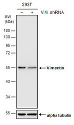
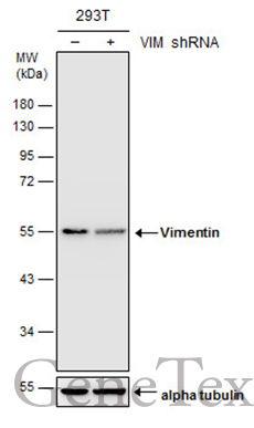
- Experimental details
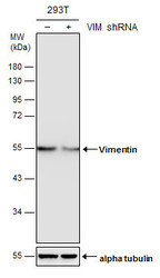
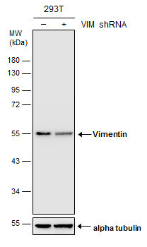
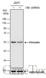
- Non-transfected (¡V) and transfected (+) 293T whole cell extracts (30 ?g) were separated by 10% SDS-PAGE, and the membrane was blotted with Vimentin antibody (GTX100619) diluted at 1:20000. The HRP-conjugated anti-rabbit IgG antibody (GTX213110-01) was used to detect the primary antibody.
Supportive validation
- Submitted by
- GeneTex (provider)
- Main image
- Experimental details
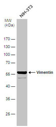
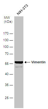
- Sample (30 ug of whole cell lysate) A:NIH-3T310% SDS PAGE GTX100619 diluted at 1:1000
- Validation comment
- WB

- Submitted by
- GeneTex (provider)
- Main image

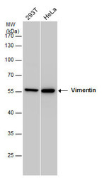
- Experimental details
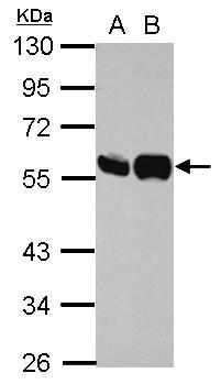
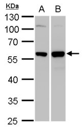
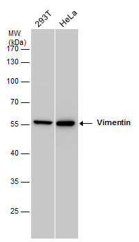
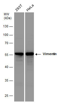
- Sample (30 ug of whole cell lysate) A: 293T B: HeLa 10% SDS PAGE GTX100619 diluted at 1:10000
- Validation comment
- WB
- Submitted by
- GeneTex (provider)
- Main image
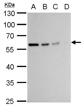
- Experimental details
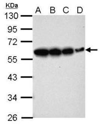
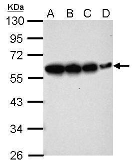
- Sample (whole cell lysate) A: 293T 20ug B: 293T 10ug C: 293T 5ug D: 293T 1ug 10% SDS PAGE GTX100619 diluted at 1:10000
- Validation comment
- WB
- Submitted by
- GeneTex (provider)
- Main image
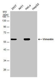
- Experimental details
- Vimentin antibody detects Vimentin protein by western blot analysis.A. 30 ?g 293T whole cell lysate/extract B. 30 ?g HeLa whole cell lysate/extract10 % SDS-PAGEVimentin antibody (GTX100619) dilution: 1:10000
- Validation comment
- WB

- Submitted by
- GeneTex (provider)
- Main image

- Experimental details
- Vimentin antibody detects Vimentin protein by western blot analysis.A. 20 ?g 293T whole cell lysate/extract B. 10 ?g 293T whole cell lysate/extract C. 5 ?g 293T whole cell lysate/extract D. 1 ?g 293T whole cell lysate/extract10% SDS-PAGEVimentin antibody (GTX100619) dilution: 1:10000The HRP-conjugated anti-rabbit IgG antibody (GTX213110-01) was used to detect the primary antibody.
- Submitted by
- GeneTex (provider)
- Main image
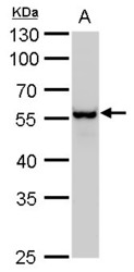
- Experimental details
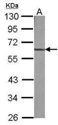
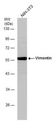
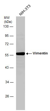
- Vimentin antibody detects Vimentin protein by western blot analysis.A. 30 ?g NIH-3T3 whole cell lysate/extract10 % SDS-PAGEVimentin antibody (GTX100619) dilution: 1:2000
- Validation comment
- WB
- Submitted by
- GeneTex (provider)
- Main image
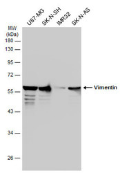
- Experimental details
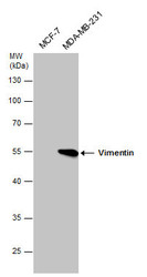
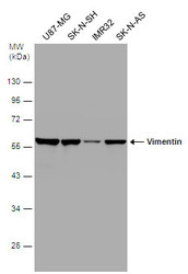
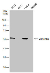
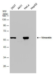
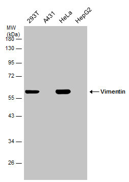
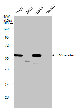
- Vimentin antibody detects Vimentin protein by western blot analysis. Various whole cell extracts (30 ?g) were separated by 10% SDS-PAGE, and the membrane was blotted with Vimentin antibody (GTX100619) diluted at a dilution of 1:10000.
- Validation comment
- WB
- Submitted by
- GeneTex (provider)
- Main image

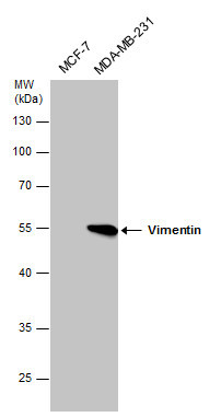
- Experimental details
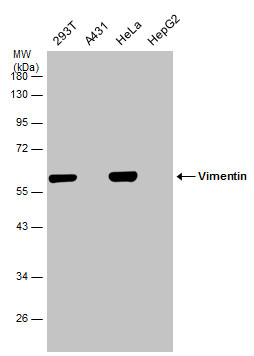
- Vimentin antibody detects Vimentin protein by Western blot analysis. Various whole cell extracts (30 £gg) were separated by 10% SDS-PAGE, and the membrane was blotted with Vimentin antibody (GTX100619) diluted at a dilution of 1:10000.
- Submitted by
- GeneTex (provider)
- Main image
- Experimental details
- Whole cell extract (30 ?g) was separated by 10% SDS-PAGE, and the membrane was blotted with Vimentin antibody (GTX100619) diluted at 1:5000. The HRP-conjugated anti-rabbit IgG antibody (GTX213110-01) was used to detect the primary antibody.
- Submitted by
- GeneTex (provider)
- Main image
- Experimental details
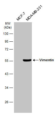
- Vimentin antibody detects Vimentin protein by western blot analysis. Whole cell extracts (30 ?g) was separated by 10% SDS-PAGE, and the membrane was blotted with Vimentin antibody (GTX100619) at a dilution of 1:10000. The HRP-conjugated anti-rabbit IgG antibody (GTX213110-01) was used to detect the primary antibody.
- Submitted by
- GeneTex (provider)
- Main image
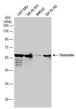
- Experimental details
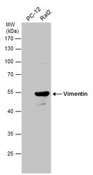
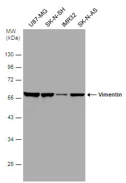
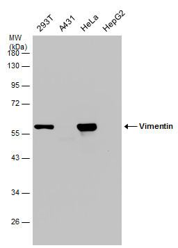
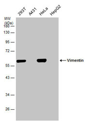
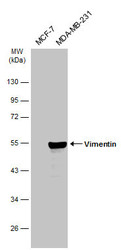
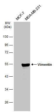
- Various whole cell extracts (30 ?g) were separated by 10% SDS-PAGE, and the membrane was blotted with Vimentin antibody (GTX100619) diluted at 1:10000. The HRP-conjugated anti-rabbit IgG antibody (GTX213110-01) was used to detect the primary antibody.
- Submitted by
- GeneTex (provider)
- Main image
- Experimental details
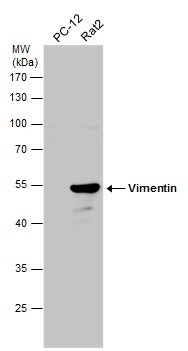
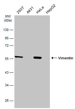
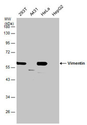
- Various whole cell extracts (30 ?g) were separated by 10% SDS-PAGE, and the membrane was blotted with Vimentin antibody (GTX100619) diluted at 1:50000. The HRP-conjugated anti-rabbit IgG antibody (GTX213110-01) was used to detect the primary antibody.
- Submitted by
- GeneTex (provider)
- Main image

- Experimental details
- Various whole cell extracts (30 ?g) were separated by 10% SDS-PAGE, and the membrane was blotted with Vimentin antibody (GTX100619) diluted at 1:50000. The HRP-conjugated anti-rabbit IgG antibody (GTX213110-01) was used to detect the primary antibody.
- Submitted by
- GeneTex (provider)
- Main image
- Experimental details
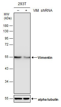
- Non-transfected (¡V) and transfected (+) 293T whole cell extracts (30 ?g) were separated by 10% SDS-PAGE, and the membrane was blotted with Vimentin antibody (GTX100619) diluted at 1:20000.
- Submitted by
- GeneTex (provider)
- Main image
- Experimental details
- Various whole cell extracts (30 ?g) were separated by 10% SDS-PAGE, and the membrane was blotted with Vimentin antibody (GTX100619) diluted at 1:50000. The HRP-conjugated anti-rabbit IgG antibody (GTX213110-01) was used to detect the primary antibody.
- Submitted by
- GeneTex (provider)
- Main image
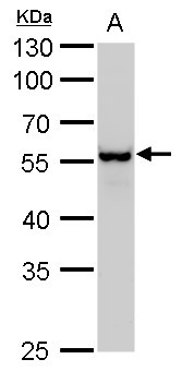
- Experimental details
- Whole cell extract (30 ?g) was separated by 10% SDS-PAGE, and the membrane was blotted with Vimentin antibody (GTX100619) diluted at 1:2000.
- Submitted by
- GeneTex (provider)
- Main image
- Experimental details
- Various whole cell extracts (30 ?g) were separated by 10% SDS-PAGE, and the membrane was blotted with Vimentin antibody (GTX100619) diluted at 1:50000. The HRP-conjugated anti-rabbit IgG antibody (GTX213110-01) was used to detect the primary antibody.
- Submitted by
- GeneTex (provider)
- Main image
- Experimental details
- Non-transfected (¡V) and transfected (+) 293T whole cell extracts (30 ?g) were separated by 10% SDS-PAGE, and the membrane was blotted with Vimentin antibody (GTX100619) diluted at 1:20000.
- Submitted by
- GeneTex (provider)
- Main image
- Experimental details
- Various whole cell extracts (30 ?g) were separated by 10% SDS-PAGE, and the membrane was blotted with Vimentin antibody (GTX100619) diluted at 1:50000. The HRP-conjugated anti-rabbit IgG antibody (GTX213110-01) was used to detect the primary antibody.
- Submitted by
- GeneTex (provider)
- Main image
- Experimental details
- Various whole cell extracts (30 ?g) were separated by 10% SDS-PAGE, and the membrane was blotted with Vimentin antibody (GTX100619) diluted at 1:50000.
- Submitted by
- GeneTex (provider)
- Main image
- Experimental details
- Various whole cell extracts (30 ?g) were separated by 10% SDS-PAGE, and the membrane was blotted with Vimentin antibody (GTX100619) diluted at 1:50000.
- Submitted by
- GeneTex (provider)
- Main image
- Experimental details
- Various whole cell extracts (30 ?g) were separated by 10% SDS-PAGE, and the membrane was blotted with Vimentin antibody (GTX100619) diluted at 1:50000.
- Submitted by
- GeneTex (provider)
- Main image
- Experimental details
- Various whole cell extracts (30 ?g) were separated by 10% SDS-PAGE, and the membrane was blotted with Vimentin antibody (GTX100619) diluted at 1:10000. The HRP-conjugated anti-rabbit IgG antibody (GTX213110-01) was used to detect the primary antibody.
Supportive validation
- Submitted by
- GeneTex (provider)
- Main image
- Experimental details
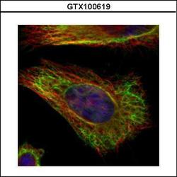
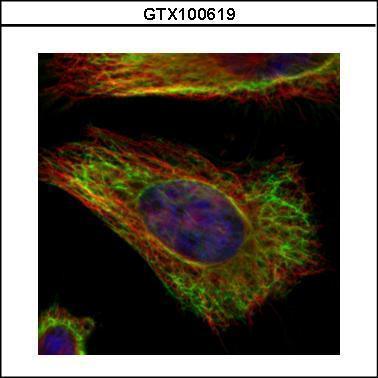
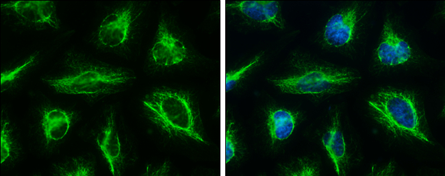
- Confocal immunofluorescence analysis (Olympus FV10i) of methanol-fixed HeLa, using Vimentin(GTX100619) antibody (Green) at 1:500 dilution. Alpha-tubulin filaments were labeled with GTX11304 (Red) at 1:2000.
- Submitted by
- GeneTex (provider)
- Main image

- Experimental details
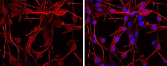
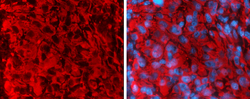
- Vimentin antibody detects Vimentin protein at cytoplasm by immunofluorescent analysis.Sample: U-87 MG cells were fixed in 4% paraformaldehyde at RT for 15 min.Red: Vimentin protein stained by Vimentin antibody (GTX100619) diluted at 1:2000.Blue: Hoechst 33342 staining.
- Submitted by
- GeneTex (provider)
- Main image
- Experimental details
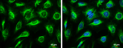
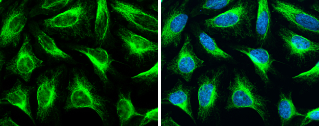
- Vimentin antibody detects Vimentin protein at cytoskeleton by immunofluorescent analysis.Sample: HeLa cells were fixed in 4% paraformaldehyde at RT for 15 min.Green: Vimentin protein stained by Vimentin antibody (GTX100619) diluted at 1:500.Blue: Hoechst 33342 staining.Scale bar = 10 £gm.

- Submitted by
- GeneTex (provider)
- Main image
- Experimental details
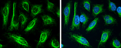
- Vimentin antibody detects Vimentin protein at cytoskeleton by immunofluorescent analysis.Sample: HeLa cells were fixed in 4% paraformaldehyde at RT for 15 min.Green: Vimentin protein stained by Vimentin antibody (GTX100619) diluted at 1:500.Blue: Hoechst 33342 staining.

- Submitted by
- GeneTex (provider)
- Main image

- Experimental details
- Vimentin antibody detects Vimentin protein at cytoskeleton by immunofluorescent analysis.Sample: HeLa cells were fixed in 4% paraformaldehyde at RT for 15 min.Green: Vimentin stained by Vimentin antibody (GTX100619) diluted at 1:500.Blue: Hoechst 33342 staining.
- Submitted by
- GeneTex (provider)
- Main image

- Experimental details
- Vimentin antibody detects Vimentin protein at cytoskeleton by immunofluorescent analysis.Sample: HeLa cells were fixed in 4% paraformaldehyde at RT for 15 min.Green: Vimentin protein stained by Vimentin antibody (GTX100619) diluted at 1:500.Blue: Hoechst 33342 staining.
Supportive validation
- Submitted by
- GeneTex (provider)
- Main image
- Experimental details
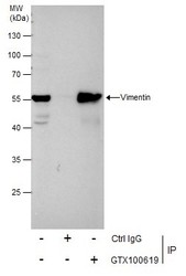
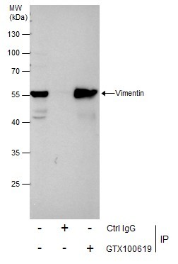
- Immunoprecipitation of Vimentin protein from HeLa whole cell extracts using 5 £gg of Vimentin antibody (GTX100619).Western blot analysis was performed using Vimentin antibody (GTX100619).EasyBlot anti-Rabbit IgG (GTX221666-01) was used as a secondary reagent.
Supportive validation
- Submitted by
- GeneTex (provider)
- Main image
- Experimental details
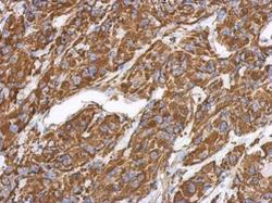
- Immunohistochemical analysis of paraffin-embedded U373 xenograft, using Vimentin(GTX100619) antibody at 1:500 dilution.

- Submitted by
- GeneTex (provider)
- Main image
- Experimental details
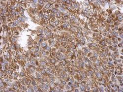
- Immunohistochemical analysis of paraffin-embedded RT2 xenograft, using Vimentin(GTX100619) antibody at 1:500 dilution.

- Submitted by
- GeneTex (provider)
- Main image

- Experimental details
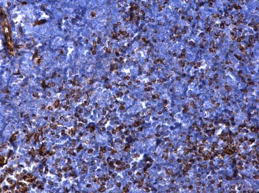
- Vimentin antibody detects Vimentin protein at cytosol on mouse thymus gland by immunohistochemical analysis. Sample: Paraffin-embedded mouse thymus gland. Vimentin antibody (GTX100619) dilution: 1:500.
- Submitted by
- GeneTex (provider)
- Main image
- Experimental details
- Immunofluorescence photomicrographs of frozen sections of mouse brain.Green: Vimentin antibody (GTX100619) diluted at 1:200. The signal was developed using goat anti-rabbit IgG antibody (Dylight594) (GTX213110-05).Blue: Nuclear staining with Hoechst 33342.

- Submitted by
- GeneTex (provider)
- Main image

- Experimental details
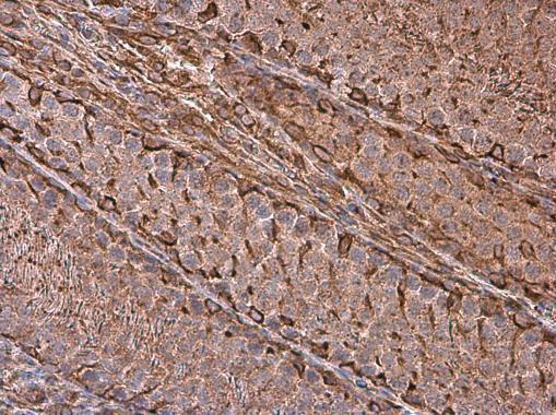
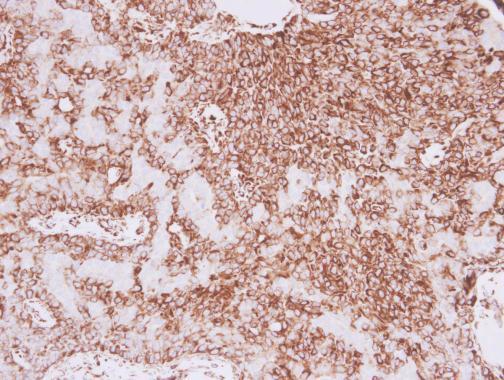
- Vimentin antibody detects Vimentin protein at cytoplasm in human lung adenocarcinoma by immunohistochemical analysis. Sample: Paraffin-embedded human lung adenocarcinoma. Vimentin antibody (GTX100619) diluted at 1:500.
- Submitted by
- GeneTex (provider)
- Main image
- Experimental details
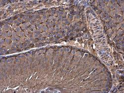
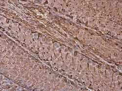
- Vimentin antibody detects Vimentin protein at cytoplasm in rat testis by immunohistochemical analysis. Sample: Paraffin-embedded rat testis. Vimentin antibody (GTX100619) diluted at 1:500.
- Submitted by
- GeneTex (provider)
- Main image
- Experimental details
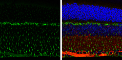
- Vimentin antibody detects Vimentin protein expression by immunohistochemical analysis.Sample:Paraffin-Embedded adult mouse retina. Green: Vimentin protein stained by Vimentin antibody (GTX100619) diluted at 1:250.Red: beta Tubulin 3/ TUJ1, stained by beta Tubulin 3/ TUJ1 antibody [GT11710] (GTX631836) diluted at 1:500.Blue: Fluoroshield with DAPI (GTX30920).

- Submitted by
- GeneTex (provider)
- Main image
- Experimental details
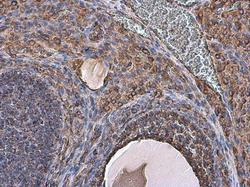
- Vimentin antibody detects Vimentin protein at cell membrane and cytoplasm in rat ovary by immunohistochemical analysis. Sample: Paraffin-embedded rat ovary. Vimentin antibody (GTX100619) diluted at 1:500.

- Submitted by
- GeneTex (provider)
- Main image
- Experimental details
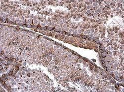
- Vimentin antibody detects Vimentin protein at cell membrane and cytoplasm in mouse testis by immunohistochemical analysis. Sample: Paraffin-embedded mouse testis. Vimentin antibody (GTX100619) diluted at 1:500.
- Submitted by
- GeneTex (provider)
- Main image
- Experimental details
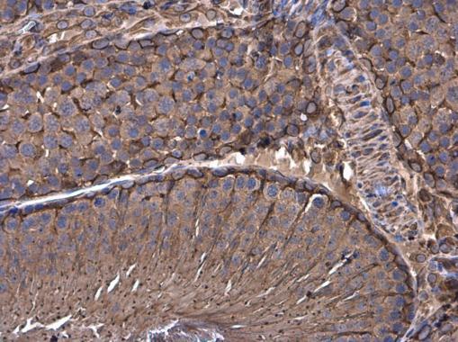
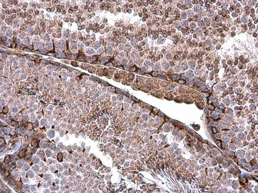
- Vimentin antibody detects Vimentin protein at cell membrane and cytoplasm in rat testis by immunohistochemical analysis. Sample: Paraffin-embedded rat testis. Vimentin antibody (GTX100619) diluted at 1:500.