 Explore
 Validate
 Learn
Learn Western blot
Antibody data
- Antibody Data
- Antigen structure
- References [0]
- Comments [0]
- Validations
- Western blot [1]
- Immunocytochemistry [1]
Submit
Validation data
Reference
Comment
Report error
- Product number
- 702180 - Provider product page

- Provider
- Invitrogen Antibodies
- Product name
- Phospho-Vimentin (Ser56) Recombinant Rabbit Monoclonal Antibody (7H18L9)
- Antibody type
- Monoclonal
- Antigen
- Synthetic peptide
- Reactivity
- Human
- Host
- Rabbit
- Isotype
- IgG
- Antibody clone number
- 7H18L9
- Vial size
- 100 µg
- Concentration
- 0.5 mg/mL
- Storage
- Store at 4°C short term. For long term storage, store at -20°C, avoiding freeze/thaw cycles.
No comments: Submit comment
Supportive validation
- Submitted by
- Invitrogen Antibodies (provider)
- Main image

- Experimental details
- Western blot analysis was performed on whole cell extracts (30 µg lysate) of HeLa (Lane 1), HeLa treated with Paclitaxel (100 nM/mL for 20hrs) (Lane 2), K-562 (Lane 3) and K-562 treated with Paclitaxel (100 nM/mL for 20hrs) (Lane 4). The blots were probed with Anti-Vimentin (pS56) Recombinant Rabbit Monoclonal Antibody (Product # 702180, 1-2 µg/mL) and detected by chemiluminescence using Goat anti-Rabbit IgG (H+L) Superclonal Secondary Antibody, HRP conjugate (Product # A27036, 0.4 µg/mL, 1:2500 dilution). A 54 kDa band corresponding to Vimentin (pS56) was observed across the cell lines tested. Known quantity of protein samples were electrophoresed using Novex®NuPAGE®4-12% Bis-Tris gel (Product # NP0321BOX), XCell SureLock Electrophoresis System (Product # EI0002) and Novex® Sharp Pre-Stained Protein Standard (Product # LC5800). Resolved proteins were then transferred onto a nitrocellulose membrane with iBlot® Dry Blotting System (Product # IB21001). The membrane was probed with the relevant primary and secondary Antibody following blocking with 5% skimmed milk. Chemiluminescent detection was performed using Pierce™ ECL Western blotting Substrate (Product # 32106).

Supportive validation
- Submitted by
- Invitrogen Antibodies (provider)
- Main image
- Experimental details
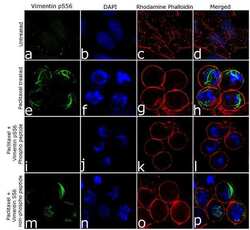
- For immunofluorescence analysis HeLa cells were fixed and permeabilized for detection of endogenous Vimentin (pS56) using Vimentin (pS56) Recombinant Rabbit Monoclonal Antibody (Product # 702180, 2 µg/mL) and labeled with Goat anti-Rabbit IgG (H+L) Superclonal Secondary Antibody, Alexa Fluor® 488 conjugate (Product # A27034, 1:2000). Nuclei (blue) were stained using SlowFade® Gold Antifade Mountant with DAPI (Product # S36938), and Alexa Fluor® 555 Rhodamine Phalloidin (Product # R415, 1:300) was used for cytoskeletal F-actin (red) staining. Detection and localization of Vimentin (pS56) (green) in the cytoplasm can be clearly observed in cells treated with paclitaxel (100 ng/mL, 20 h) as compared to untreated cells. Antibody specificity was demonstrated by competition with the Vimentin (pS56) peptide, which results in loss of signal. No competition was observed with the non-phospho peptide. The images were captured at 60X magnification. The antibody detected a faint nuclear signal for Vimentin pS56 in untreated cells, which was also competed out specifically by the phospho peptide (data not shown). Although vimentin is predominantly cytoplasmic, its nuclear localization has been reported. The role of S56 phosphorylation in the nucleus however has not been established to date.
