 Explore
 Validate
 Learn
Learn Immunocytochemistry
Antibody data
- Antibody Data
- Antigen structure
- References [8]
- Comments [0]
- Validations
- Immunocytochemistry [2]
- Other assay [6]
Submit
Validation data
Reference
Comment
Report error
- Product number
- 11-9897-80 - Provider product page

- Provider
- Invitrogen Antibodies
- Product name
- Vimentin Monoclonal Antibody (V9), FITC, eBioscience™
- Antibody type
- Monoclonal
- Antigen
- Other
- Description
- Description: The V9 monoclonal antibody recognizes human Vimentin, a 57 kDa protein that functions as a structural component of intermediate filaments. Vimentin is expressed in cells derived from the mesenchyme but also in specific populations such as radial glia and immature glial cells, pancreatic precursor cells. It is proposed to be a marker of cardiac differentiation. In neural cells, vimentin expression is gradually replaced by neurofilaments. Reports have also shown surface expression of vimentin on activated macrophages, platelets, as well as apoptotic T cells and neutrophils.
- Conjugate
- Green dye
- Antibody clone number
- V9
- Concentration
- 0.5 mg/mL
Submitted references Characterization of epithelial cells, connective tissue cells and immune cells in human upper airway mucosa by immunofluorescence multichannel image cytometry: a pilot study.
Defining tumor-associated vascular heterogeneity in pediatric high-grade and diffuse midline gliomas.
MicroRNA-384 inhibits nasopharyngeal carcinoma growth and metastasis via binding to Smad5 and suppressing the Wnt/β-catenin axis.
Omentin-1 is associated with atrial fibrillation in patients with cardiac valve disease.
GMP-compatible and xeno-free cultivation of mesenchymal progenitors derived from human-induced pluripotent stem cells.
Levels of hepatic Th17 cells and regulatory T cells upregulated by hepatic stellate cells in advanced HBV-related liver fibrosis.
Chronic macrophage migration inhibitory factor exposure induces mesenchymal epithelial transition and promotes gastric and colon cancers.
Expression of the progenitor marker NG2/CSPG4 predicts poor survival and resistance to ionising radiation in glioblastoma.
Giotakis AI, Dudas J, Glueckert R, Dejaco D, Ingruber J, Fleischer F, Innerhofer V, Pinggera L, Bektic-Tadic L, Gabriel SAM, Riechelmann H
Histochemistry and cell biology 2021 Mar;155(3):405-421
Histochemistry and cell biology 2021 Mar;155(3):405-421
Defining tumor-associated vascular heterogeneity in pediatric high-grade and diffuse midline gliomas.
Wei X, Meel MH, Breur M, Bugiani M, Hulleman E, Phoenix TN
Acta neuropathologica communications 2021 Aug 23;9(1):142
Acta neuropathologica communications 2021 Aug 23;9(1):142
MicroRNA-384 inhibits nasopharyngeal carcinoma growth and metastasis via binding to Smad5 and suppressing the Wnt/β-catenin axis.
Zeng X, Liao H, Wang F
Cytotechnology 2021 Apr;73(2):203-215
Cytotechnology 2021 Apr;73(2):203-215
Omentin-1 is associated with atrial fibrillation in patients with cardiac valve disease.
Chen Y, Liu F, Han F, Lv L, Tang CE, Xie Z, Luo F
BMC cardiovascular disorders 2020 May 6;20(1):214
BMC cardiovascular disorders 2020 May 6;20(1):214
GMP-compatible and xeno-free cultivation of mesenchymal progenitors derived from human-induced pluripotent stem cells.
McGrath M, Tam E, Sladkova M, AlManaie A, Zimmer M, de Peppo GM
Stem cell research & therapy 2019 Jan 11;10(1):11
Stem cell research & therapy 2019 Jan 11;10(1):11
Levels of hepatic Th17 cells and regulatory T cells upregulated by hepatic stellate cells in advanced HBV-related liver fibrosis.
Li X, Su Y, Hua X, Xie C, Liu J, Huang Y, Zhou L, Zhang M, Li X, Gao Z
Journal of translational medicine 2017 Apr 11;15(1):75
Journal of translational medicine 2017 Apr 11;15(1):75
Chronic macrophage migration inhibitory factor exposure induces mesenchymal epithelial transition and promotes gastric and colon cancers.
Morris KT, Nofchissey RA, Pinchuk IV, Beswick EJ
PloS one 2014;9(6):e98656
PloS one 2014;9(6):e98656
Expression of the progenitor marker NG2/CSPG4 predicts poor survival and resistance to ionising radiation in glioblastoma.
Svendsen A, Verhoeff JJ, Immervoll H, Brøgger JC, Kmiecik J, Poli A, Netland IA, Prestegarden L, Planagumà J, Torsvik A, Kjersem AB, Sakariassen PØ, Heggdal JI, Van Furth WR, Bjerkvig R, Lund-Johansen M, Enger PØ, Felsberg J, Brons NH, Tronstad KJ, Waha A, Chekenya M
Acta neuropathologica 2011 Oct;122(4):495-510
Acta neuropathologica 2011 Oct;122(4):495-510
No comments: Submit comment
Supportive validation
- Submitted by
- Invitrogen Antibodies (provider)
- Main image

- Experimental details
- Immunocytochemistry of fixed and permeabilized C6 cells using 1 µg/mL of Mouse IgG1 Isotype Control FITC (Product # 11-4714-42) (left) or 1 µg/mL of Anti-Vimentin FITC (right). Nuclei are counterstained with DAPI.
- Conjugate
- Green dye

Supportive validation
- Submitted by
- Invitrogen Antibodies (provider)
- Main image

- Experimental details
- Fig. 2 Supernatants from HSC increased the percentages of Th17 cells and Tregs. a Phenotypes of primary HSC extracted from HBV-related fibrotic liver tissues in Group 2. Sections were immunostained with desmin, FAP, FSP, vimentin, fibronectin and a-SMA antibodies. One of 20 representative micrographs is shown. b , d Purified CD4+ T cells were cultured alone (Blank) or with 30% indicated supernatants. The values in the quadrants represent the percentages of Th17 cells and Tregs. The data shown are representative dot plots from more than three independent experiments. c , e The statistical analysis of the effect of LX-2 and pHSC supernatant on the percentages of Th17 cells ( c ) and Tregs ( e ). * p < 0.05, ** p < 0.01, *** p < 0.001
- Conjugate
- Green dye

- Submitted by
- Invitrogen Antibodies (provider)
- Main image

- Experimental details
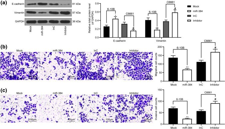
- Fig. 3 miR-384 mimic inhibits EMT, migration and invasion of NPC cells. a levels of mesenchyme marker protein Vimentin and epithelium-marker protein E-cadherin in cells measured by western blot analysis; b , c number of migrated ( b ) and invaded ( c ) cells determined by Transwell assays. Data are exhibited as mean +- SD from three independent experiments; in panel ( a ), data were analyzed using two-way ANOVA, while data in panels ( b ) and ( c ) were analyzed by one-way ANOVA, and Tukey''s multiple comparison test was used for the post-hoc test after ANOVA; * p < 0.05; ** p < 0.01 vs. the Mock group; # p < 0.05 vs. the InC group
- Conjugate
- Green dye
- Submitted by
- Invitrogen Antibodies (provider)
- Main image

- Experimental details
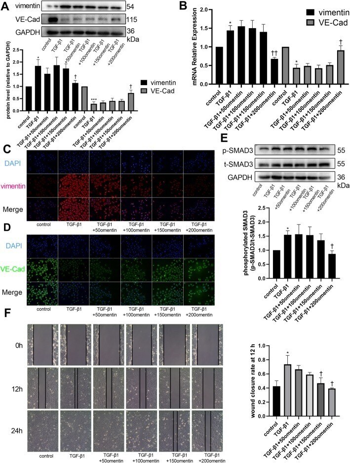
- Fig. 3 Omentin-1 inhibited TGF-beta1-induced endothelial-mesenchymal transition (EndMT) of human umbilical vein endothelial cells (HUVECs). Vimentin and VE-Cad protein levels in HUVECs were detected via western blotting ( a ). The mRNA levels of vimentin and VE-Cad were detected via RT-qPCR ( b ). Expression of mRNA were normalized to GAPDH. Representative images of immunofluorescence staining for vimentin ( c ) and VE-Cad ( d ) (200x magnification) in HUVECs. The p-SMAD3 and t-SMAD3 protein levels in HUVECs were detected via western blotting ( e ). ( f ) Representative images of HUVECs scratch assay (x 100 magnification). The values were mean +- SEM of three independent experiments. * P < 0.05 vs control group, ** P < 0.01 vs control group, *** P < 0.001 vs control group, + P < 0.05 vs TGF-beta1 group, ++ P < 0.01 vs TGF-beta1 group, +++ P < 0.001 vs TGF-beta1 group
- Conjugate
- Green dye
- Submitted by
- Invitrogen Antibodies (provider)
- Main image

- Experimental details
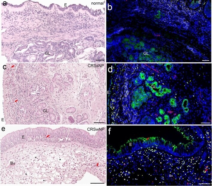
- Fig. 3 Chronic rhinosinusitis types and normal tissue; comparison of bright-field and fluorescent-immunolabeled sections. H.E. staining (a, c, e) and immunofluorescence staining (b, d, f) of control (a, b) , chronic rhinosinusitis without polyps (CRSsNP) (c, d) and chronic rhinosinusitis with polyps (CRSwNP) (e, f) tissue. Four-channel fluorescence represents nuclear DAPI staining (blue), cytokeratin (green), vimentin (red) and CD45 combined with CD18 (yellow). Scale bar: 100 mum
- Conjugate
- Green dye
- Submitted by
- Invitrogen Antibodies (provider)
- Main image

- Experimental details
- Fig. 4 Comparison of histograms of fluorescence signals between isotype controls and patient's tissue. Histograms of fluorescence signals after control incubation in the upper histograms (isotype controls) and specific fluorescence signals after test incubation in the lower histograms (patient's tissue). Presentation of a random cell sample of all patients and their isotype controls. X -axis: mean intensity in logarithmic scale. Y -axis: cell count. The left histograms represented cytokeratin fluorescence signal, the middle histograms vimentin fluorescence signal and the right histograms CD45/CD18 fluorescence signal
- Conjugate
- Green dye

- Submitted by
- Invitrogen Antibodies (provider)
- Main image

- Experimental details
- Fig. 5 Cytoskeletal proteins. Cell painting showing the production of the cytoskeletal proteins F-actin and vimentin in induced pluripotent stem cell-derived mesodermal progenitors (line 1013A and BC1) cultured in different media at passages 6, 10, and 15. The nuclei are stained with 4',6-diamidino-2-phenylindole (blue). Scale bar = 20 mum. Abbreviations: MP, mesodermal progenitors; FBS, fetal bovine serum; HPL, human platelet lysate; P, passage. Additional data are shown in Additional file 4 : Figure S3
- Conjugate
- Green dye
