Explore
 Validate
 Learn
Learn Western blot
Antibody data
- Antibody Data
- Antigen structure
- References [0]
- Comments [0]
- Validations
- Western blot [1]
- Immunocytochemistry [2]
- Immunohistochemistry [1]
- Other assay [1]
Submit
Validation data
Reference
Comment
Report error
- Product number
- MAB21052-100 - Provider product page

- Provider
- R&D Systems
- Product name
- Human/Mouse/Rat Vimentin Antibody
- Antibody type
- Monoclonal
- Description
- Protein A or G purified from hybridoma culture supernatant. Detects human Vimentin in direct ELISAs. Detects human, mouse, and rat Vimentin in Western blots.
- Reactivity
- Human, Mouse, Rat
- Host
- Mouse
- Conjugate
- Unconjugated
- Antigen sequence
P08670- Isotype
- IgG
- Antibody clone number
- 979517
- Vial size
- 100 ug
- Storage
- Use a manual defrost freezer and avoid repeated freeze-thaw cycles. 12 months from date of receipt, -20 to -70 °C as supplied. 1 month, 2 to 8 °C under sterile conditions after reconstitution. 6 months, -20 to -70 °C under sterile conditions after reconstitution.
No comments: Submit comment
Supportive validation
- Submitted by
- R&D Systems (provider)
- Main image

- Experimental details
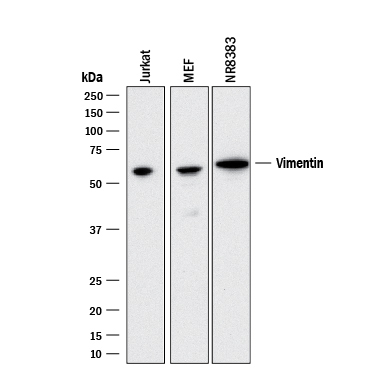
- Detection of Human, Mouse, and Rat Vimentin by Western Blot. Western blot shows lysates of Jurkat human acute T cell leukemia cell line, MEF mouse embryonic feeder cells, and NR8383 rat alveolar macrophage cell line. PVDF membrane was probed with 2 µg/mL of Mouse Anti-Human/Mouse/Rat Vimentin Monoclonal Antibody (Catalog # MAB21052) followed by HRP-conjugated Anti-Mouse IgG Secondary Antibody (Catalog # HAF018). A specific band was detected for Vimentin at approximately 55 kDa (as indicated). This experiment was conducted under reducing conditions and using Immunoblot Buffer Group 3.
Supportive validation
- Submitted by
- R&D Systems (provider)
- Main image

- Experimental details
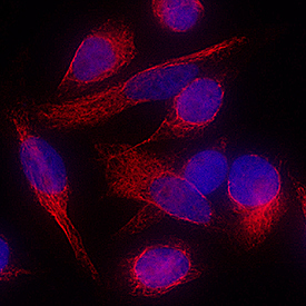
- Vimentin in HeLa Human Cell Line. Vimentin was detected in immersion fixed HeLa human cervical epithelial carcinoma cell line using Mouse Anti-Human/Mouse/Rat Vimentin Monoclonal Antibody (Catalog # MAB21052) at 8 µg/mL for 3 hours at room temperature. Cells were stained using the NorthernLights™ 557-conjugated Anti-Mouse IgG Secondary Antibody (red; Catalog # NL007) and counterstained with DAPI (blue). Specific staining was localized to cytoplasm and cytoskeleton. View our protocol for Fluorescent ICC Staining of Cells on Coverslips.
- Submitted by
- R&D Systems (provider)
- Main image

- Experimental details
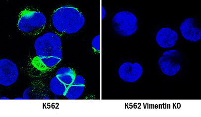
- Vimentin Specificity is Shown by Immunocytochemistry in Knockout Cell Line. Vimentin was detected in immersion fixed K562 human chronic myelogenous leukemia cell line but is not detected in Vimentin knockout (KO) K562 Human Cell Line cell line using Mouse Anti-Human/Mouse/Rat Vimentin Monoclonal Antibody (Catalog # MAB21052) at 5 µg/mL for 3 hours at room temperature. Cells were stained using the NorthernLights 493-conjugated Anti-Mouse IgG Secondary Antibody (green; Catalog # NL009) and counterstained with DAPI (blue). Specific staining was localized to cytoplasm. View our protocol for Fluorescent ICC Staining of Non-adherent Cells.
Supportive validation
- Submitted by
- R&D Systems (provider)
- Main image
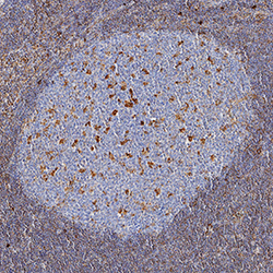
- Experimental details
- Vimentin in Human Lymph Node. Vimentin was detected in immersion fixed paraffin-embedded sections of human lymph node using Mouse Anti-Human/Mouse/Rat Vimentin Monoclonal Antibody (Catalog # MAB21052) at 5 µg/mL for 1 hour at room temperature followed by incubation with the Anti-Mouse IgG VisUCyte™ HRP Polymer Antibody (Catalog # VC001). Tissue was stained using DAB (brown) and counterstained with hematoxylin (blue). Specific staining was localized to cytoplasm in lymphocytes. View our protocol for IHC Staining with VisUCyte HRP Polymer Detection Reagents.

Supportive validation
- Submitted by
- R&D Systems (provider)
- Main image
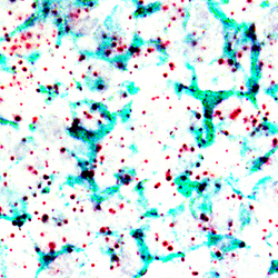
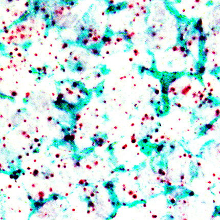
- Experimental details
- Vimentin in Human Tonsil Using Dual RNAscope® ISH and IHC. Vimentin mRNA (red) and protein (green) was detected in formalin-fixed paraffin-embedded tissue sections of human tonsil probed with ACD RNAScope® Probe (Catalog # 310441) followed by immunohistochemistry using R&D Systems Mouse Anti-Human/Mouse/Rat Vimentin Monoclonal Antibody (Catalog# MAB21052) at 5ug/mL for 1 hour at room temperature followed by incubation with the Anti-Mouse IgG VisUCyte HRP Polymer Antibody (R&D Systems, Catalog # VC001). Tissue was stained using ACD RNAscope® 2.5 HD Duplex Detection Reagents (Catalog # 322500).