Explore
 Validate
 Learn
Learn Western blot
Antibody data
- Antibody Data
- Antigen structure
- References [0]
- Comments [0]
- Validations
- Western blot [5]
- Immunocytochemistry [1]
- Immunohistochemistry [16]
Submit
Validation data
Reference
Comment
Report error
- Product number
- CF801297 - Provider product page

- Provider
- Invitrogen Antibodies
- Product name
- VIM Monoclonal Antibody (OTI1A9), TrueMAB™
- Antibody type
- Monoclonal
- Antigen
- Recombinant full-length protein
- Reactivity
- Human
- Host
- Mouse
- Isotype
- IgG
- Antibody clone number
- OTI1A9
- Vial size
- 100 µg
- Concentration
- 1 mg/mL
- Storage
- -20° C, Avoid Freeze/Thaw Cycles
No comments: Submit comment
Supportive validation
- Submitted by
- Invitrogen Antibodies (provider)
- Main image
- Experimental details
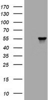
- HEK293T cells were transfected with the pCMV6-ENTRY control (Left lane) or pCMV6-ENTRY VIM (RC201546, Right lane) cDNA for 48 hrs and lysed. Equivalent amounts of cell lysates (5 µg per lane) were separated by SDS-PAGE and immunoblotted with anti-VIM. (1:2. Positive lysates LY401165 (100 µg) and LC401165 (20 µg) can be purchased separately from OriGene.
- Submitted by
- Invitrogen Antibodies (provider)
- Main image
- Experimental details
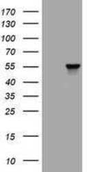
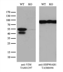
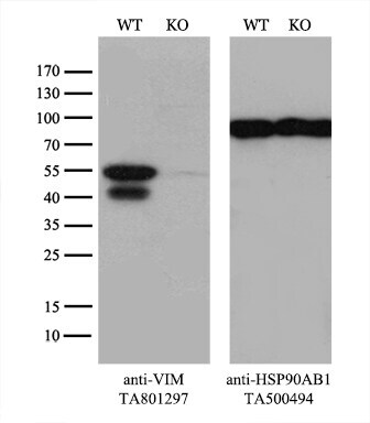
- Equivalent amounts of cell lysates (10 µg per lane) of wild-type Hela cells (WT, Cat# LC810HELA) and VIM-Knockout Hela cells (KO, Cat# LC810257) were separated by SDS-PAGE and immunoblotted with anti-VIM monoclonal antibody TA801297, (1:500). Then the blotted membrane was stripped and reprobed with anti-HSP90AB1 antibody (TA500494) as a loading control.
- Submitted by
- Invitrogen Antibodies (provider)
- Main image
- Experimental details
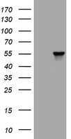
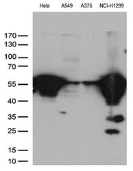
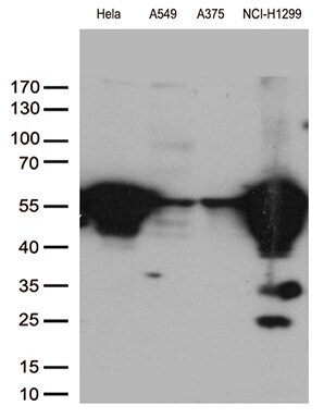
- Western blot analysis of extracts (35 µg) from 4 different cell lines by using anti-VIM monoclonal antibody. (1:500)
- Submitted by
- Invitrogen Antibodies (provider)
- Main image

- Experimental details
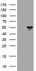
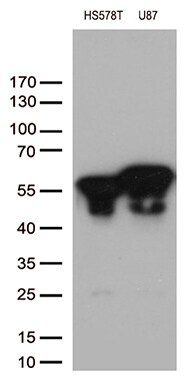
- Western blot analysis of extracts (35 µg) from 2 different cell lines by using anti-VIM monoclonal antibody. (1:500)
Supportive validation
- Submitted by
- Invitrogen Antibodies (provider)
- Main image
- Experimental details
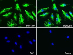
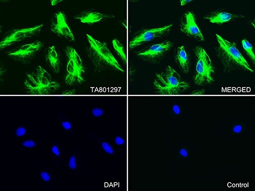
- Immunofluorescent staining of Hela cells using anti-VIM mouse monoclonal antibody (TA801297, green, upper left; merged, upper right) or Isotype control (merged, lower right). Cell nuclei were stained with DAPI (blue, lower left). (1:100)
Supportive validation
- Submitted by
- Invitrogen Antibodies (provider)
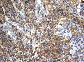
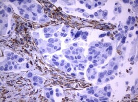
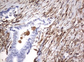
- Main image

- Experimental details
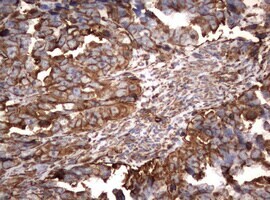
- Immunohistochemical staining of paraffin-embedded Carcinoma of Human lung tissue using anti-VIM mouse monoclonal antibody. (Heat-induced epitope retrieval by 10mM citric buffer, pH6.0, 120°C for 3min, TA801297)(1:150)
- Submitted by
- Invitrogen Antibodies (provider)
- Main image
- Experimental details
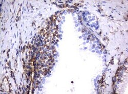
- Immunohistochemical staining of paraffin-embedded human Ovary tissue within the normal limits using anti-VIM mouse monoclonal antibody. (Heat-induced epitope retrieval by 10mM citric buffer, pH6.0, 120°C for 3min, TA801297)(1:150)

- Submitted by
- Invitrogen Antibodies (provider)
- Main image
- Experimental details
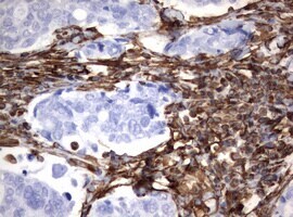
- Immunohistochemical staining of paraffin-embedded Adenocarcinoma of Human ovary tissue using anti-VIM mouse monoclonal antibody. (Heat-induced epitope retrieval by 10mM citric buffer, pH6.0, 120°C for 3min, TA801297)(1:150)
- Submitted by
- Invitrogen Antibodies (provider)
- Main image
- Experimental details
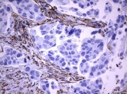
- Immunohistochemical staining of paraffin-embedded Carcinoma of Human thyroid tissue using anti-VIM mouse monoclonal antibody. (Heat-induced epitope retrieval by 10mM citric buffer, pH6.0, 120°C for 3min, TA801297)(1:150)
- Submitted by
- Invitrogen Antibodies (provider)
- Main image
- Experimental details
- Immunohistochemical staining of paraffin-embedded human endometrium tissue within the normal limits using anti-VIM mouse monoclonal antibody. (Heat-induced epitope retrieval by 10mM citric buffer, pH6.0, 120°C for 3min, TA801297)(1:150)

- Submitted by
- Invitrogen Antibodies (provider)
- Main image
- Experimental details
- Immunohistochemical staining of paraffin-embedded Adenocarcinoma of Human endometrium tissue using anti-VIM mouse monoclonal antibody. (Heat-induced epitope retrieval by 10mM citric buffer, pH6.0, 120°C for 3min, TA801297)(1:150)
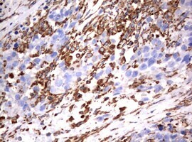
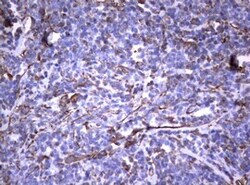
- Submitted by
- Invitrogen Antibodies (provider)
- Main image

- Experimental details
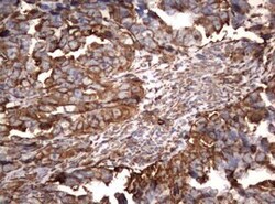
- Immunohistochemical staining of paraffin-embedded Adenocarcinoma of Human breast tissue using anti-VIM mouse monoclonal antibody. (Heat-induced epitope retrieval by 10mM citric buffer, pH6.0, 120°C for 3min, TA801297)(1:150)
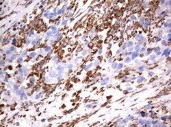
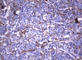
- Submitted by
- Invitrogen Antibodies (provider)
- Main image
- Experimental details
- Immunohistochemical staining of paraffin-embedded Carcinoma of Human prostate tissue using anti-VIM mouse monoclonal antibody. (Heat-induced epitope retrieval by 10mM citric buffer, pH6.0, 120°C for 3min, TA801297)(1:150)

- Submitted by
- Invitrogen Antibodies (provider)
- Main image
- Experimental details
- Immunohistochemical staining of paraffin-embedded Carcinoma of Human bladder tissue using anti-VIM mouse monoclonal antibody. (Heat-induced epitope retrieval by 10mM citric buffer, pH6.0, 120°C for 3min, TA801297)(1:150)
- Submitted by
- Invitrogen Antibodies (provider)
- Main image
- Experimental details
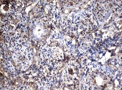
- Immunohistochemical staining of paraffin-embedded human lymph node tissue within the normal limits using anti-VIM mouse monoclonal antibody. (Heat-induced epitope retrieval by 10mM citric buffer, pH6.0, 120°C for 3min, TA801297)(1:150)

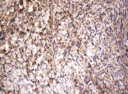
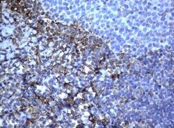
- Submitted by
- Invitrogen Antibodies (provider)
- Main image
- Experimental details
- Immunohistochemical staining of paraffin-embedded human lymphoma tissue using anti-VIM mouse monoclonal antibody. (Heat-induced epitope retrieval by 10mM citric buffer, pH6.0, 120°C for 3min, TA801297)(1:150)
- Submitted by
- Invitrogen Antibodies (provider)
- Main image

- Experimental details
- Immunohistochemical staining of paraffin-embedded Adenocarcinoma of Human colon tissue using anti-VIM mouse monoclonal antibody. (Heat-induced epitope retrieval by 10mM citric buffer, pH6.0, 120°C for 3min, TA801297)(1:150)
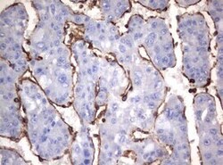
- Submitted by
- Invitrogen Antibodies (provider)
- Main image
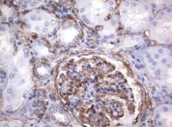
- Experimental details
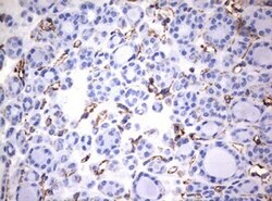
- Immunohistochemical staining of paraffin-embedded human Kidney tissue within the normal limits using anti-VIM mouse monoclonal antibody. (Heat-induced epitope retrieval by 10mM citric buffer, pH6.0, 120°C for 3min, TA801297)(1:150)

- Submitted by
- Invitrogen Antibodies (provider)
- Main image
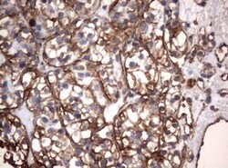
- Experimental details
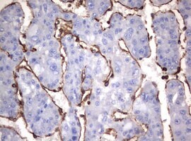
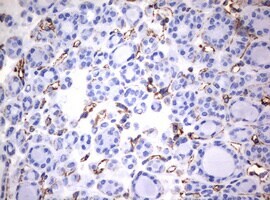
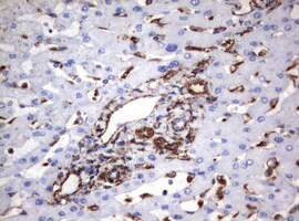
- Immunohistochemical staining of paraffin-embedded Carcinoma of Human kidney tissue using anti-VIM mouse monoclonal antibody. (Heat-induced epitope retrieval by 10mM citric buffer, pH6.0, 120°C for 3min, TA801297)(1:150)
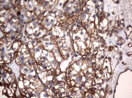
- Submitted by
- Invitrogen Antibodies (provider)
- Main image

- Experimental details
- Immunohistochemical staining of paraffin-embedded human liver tissue within the normal limits using anti-VIM mouse monoclonal antibody. (Heat-induced epitope retrieval by 10mM citric buffer, pH6.0, 120°C for 3min, TA801297)(1:150)
- Submitted by
- Invitrogen Antibodies (provider)
- Main image
- Experimental details
- Immunohistochemical staining of paraffin-embedded Carcinoma of Human liver tissue using anti-VIM mouse monoclonal antibody. (Heat-induced epitope retrieval by 10mM citric buffer, pH6.0, 120°C for 3min, TA801297)(1:150)