Explore
 Validate
 Learn
Learn Western blot
 Immunocytochemistry
Antibody data
- Antibody Data
- Antigen structure
- References [3]
- Comments [0]
- Validations
- Western blot [5]
- Immunohistochemistry [6]
Submit
Validation data
Reference
Comment
Report error
- Product number
- NBP1-85814 - Provider product page

- Provider
- Novus Biologicals
- Proper citation
- Novus Cat#NBP1-85814, RRID:AB_11020836
- Product name
- Rabbit Polyclonal Vimentin Antibody
- Antibody type
- Polyclonal
- Description
- Immunogen affinity purified. Specificity of human Vimentin antibody verified on a Protein Array containing target protein plus 383 other non-specific proteins.
- Reactivity
- Human, Mouse
- Host
- Rabbit
- Isotype
- IgG
- Vial size
- 0.1 ml
- Storage
- Store at 4C short term. Aliquot and store at -20C long term. Avoid freeze-thaw cycles.
Submitted references Changes in cannabinoid receptors, aquaporin 4 and vimentin expression after traumatic brain injury in adolescent male mice. Association with edema and neurological deficit.
Tumor suppressive microRNA‑138 contributes to cell migration and invasion through its targeting of vimentin in renal cell carcinoma.
Systematic validation of antibody binding and protein subcellular localization using siRNA and confocal microscopy.
Lopez-Rodriguez AB, Acaz-Fonseca E, Viveros MP, Garcia-Segura LM
PloS one 2015;10(6):e0128782
PloS one 2015;10(6):e0128782
Tumor suppressive microRNA‑138 contributes to cell migration and invasion through its targeting of vimentin in renal cell carcinoma.
Yamasaki T, Seki N, Yamada Y, Yoshino H, Hidaka H, Chiyomaru T, Nohata N, Kinoshita T, Nakagawa M, Enokida H
International journal of oncology 2012 Sep;41(3):805-17
International journal of oncology 2012 Sep;41(3):805-17
Systematic validation of antibody binding and protein subcellular localization using siRNA and confocal microscopy.
Stadler C, Hjelmare M, Neumann B, Jonasson K, Pepperkok R, Uhlén M, Lundberg E
Journal of proteomics 2012 Apr 3;75(7):2236-51
Journal of proteomics 2012 Apr 3;75(7):2236-51
No comments: Submit comment
Supportive validation
- Submitted by
- Novus Biologicals (provider)
- Main image

- Experimental details
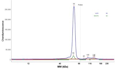
- Simple Western: Vimentin Antibody [NBP1-85814] - Simple Western lane view shows a specific band for VIM in 0.2 mg/ml of U-251MG lysate. This experiment was performed under reducing conditions using the 12-230 kDa separation system.

- Submitted by
- Novus Biologicals (provider)
- Main image

- Experimental details
- Simple Western: Vimentin Antibody [NBP1-85814] - Simple Western lane view shows a specific band for VIM in 0.2 mg/ml of NIH-3T3 lysate. This experiment was performed under reducing conditions using the 12-230 kDa separation system.

- Submitted by
- Novus Biologicals (provider)
- Main image
- Experimental details
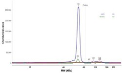
- Simple Western: Vimentin Antibody [NBP1-85814] - Electropherogram image(s) of corresponding Simple Western lane view. Vimentin antibody was used at 1:20 dilution on NIH-3T3 lysates(s).
- Submitted by
- Novus Biologicals (provider)
- Main image

- Experimental details
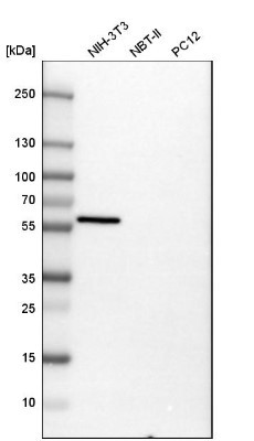
- Western Blot: Vimentin Antibody [NBP1-85814] - Western blot analysis in mouse cell line NIH-3T3, rat cell line NBT-II and rat cell line pC12.
- Submitted by
- Novus Biologicals (provider)
- Main image
- Experimental details
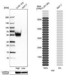
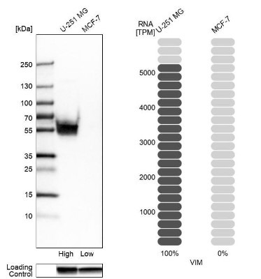
- Western Blot: Vimentin Antibody [NBP1-85814] - Analysis in human cell lines U-251MG and MCF-7 using anti-VIM antibody. Corresponding VIM RNA-seq data are presented for the same cell lines. Loading control: anti-GAPDH.
Supportive validation
- Submitted by
- Novus Biologicals (provider)
- Main image
- Experimental details
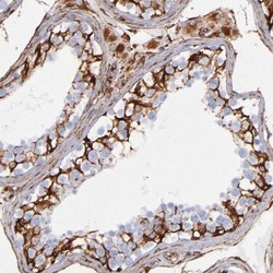
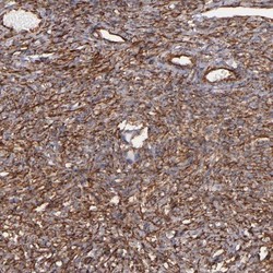
- Immunohistochemistry-Paraffin: Vimentin Antibody [NBP1-85814] - Staining of human testis shows strong membranous positivity in cells in seminiferous ducts.
- Submitted by
- Novus Biologicals (provider)
- Main image
- Experimental details
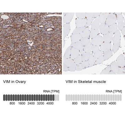
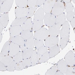
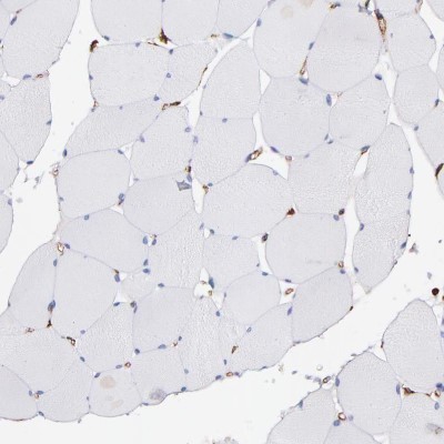
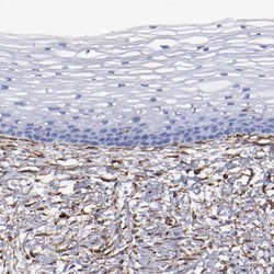
- Immunohistochemistry-Paraffin: Vimentin Antibody [NBP1-85814] - Staining of human skeletal muscle shows no positivity in myocytes.
- Submitted by
- Novus Biologicals (provider)
- Main image
- Experimental details
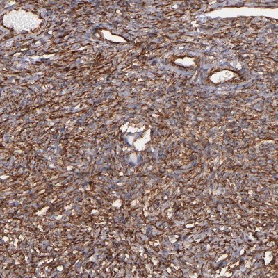
- Immunohistochemistry-Paraffin: Vimentin Antibody [NBP1-85814] - Staining of human ovary shows moderate cytoplasmic positivity in stromal cells.
- Submitted by
- Novus Biologicals (provider)
- Main image

- Experimental details
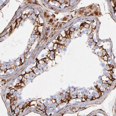
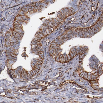
- Immunohistochemistry-Paraffin: Vimentin Antibody [NBP1-85814] - Staining of human fallopian tube shows moderate membranous positivity in glandular cells.
- Submitted by
- Novus Biologicals (provider)
- Main image
- Experimental details
- Immunohistochemistry-Paraffin: Vimentin Antibody [NBP1-85814] - Staining of human cervix, uterine shows moderate cytoplasmic positivity in stromal cells.

- Submitted by
- Novus Biologicals (provider)
- Main image

- Experimental details
- Immunohistochemistry-Paraffin: Vimentin Antibody [NBP1-85814] - Analysis in human ovary and skeletal muscle tissues. Corresponding VIM RNA-seq data are presented for the same tissues.