 Explore
 Validate
 Learn
Learn Western blot
 ELISA
ELISAAntibody data
- Antibody Data
- Antigen structure
- References [19]
- Comments [0]
- Validations
- ELISA [3]
- Immunocytochemistry [6]
- Immunohistochemistry [9]
- Other assay [15]
Submit
Validation data
Reference
Comment
Report error
- Product number
- PA5-27231 - Provider product page

- Provider
- Invitrogen Antibodies
- Product name
- Vimentin Polyclonal Antibody
- Antibody type
- Polyclonal
- Antigen
- Recombinant full-length protein
- Description
- Recommended positive controls: 293T, HeLa, MDA-MB-231, U87-MG, SK-N-SH, IMR32, SK-N-AS, NIH-3T3, A549. Predicted reactivity: Mouse (98%), Rat (98%), Xenopus laevis (85%), Dog (98%), Chicken (92%), Chimpanzee (99%), Bovine (98%), Guinea pig (97%). Store product as a concentrated solution. Centrifuge briefly prior to opening the vial.
- Reactivity
- Human, Mouse, Rat
- Host
- Rabbit
- Isotype
- IgG
- Vial size
- 100 μL
- Concentration
- 0.58 mg/mL
- Storage
- Store at 4°C short term. For long term storage, store at -20°C, avoiding freeze/thaw cycles.
Submitted references Comparative analysis of ChAdOx1 nCoV-19 and Ad26.COV2.S SARS-CoV-2 vector vaccines.
Identification of the Molecular Basis of Nanocurcumin-Induced Telocyte Preservation within the Colon of Ulcerative Colitis Rat Model.
E2F5 Promotes the Malignancy of Ovarian Cancer Via the Regulation of Hippo and Wnt Pathways.
CD44 alternative splicing senses intragenic DNA methylation in tumors via direct and indirect mechanisms.
D-Propranolol Impairs EGFR Trafficking and Destabilizes Mutant p53 Counteracting AKT Signaling and Tumor Malignancy.
Establishment and Characterization of a Novel Dedifferentiated Chondrosarcoma Cell Line DDCS2.
Ginsenoside Rg3 inhibits osteosarcoma progression by reducing circ_0003074 expression in a miR-516b-5p/KPNA4-dependent manner.
Mitotic Activation Around Wound Edges and Epithelialization Repair in UVB-Induced Capsular Cataracts.
Differential expression and inhibitory effects of aquaporins on the development of adenomyosis.
FAT4 silencing promotes epithelial-to-mesenchymal transition and invasion via regulation of YAP and β-catenin activity in ovarian cancer.
Periconceptional alcohol exposure causes female-specific perturbations to trophoblast differentiation and placental formation in the rat.
Citrullination Controls Dendritic Cell Transdifferentiation into Osteoclasts.
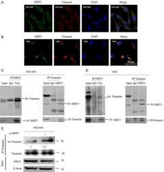
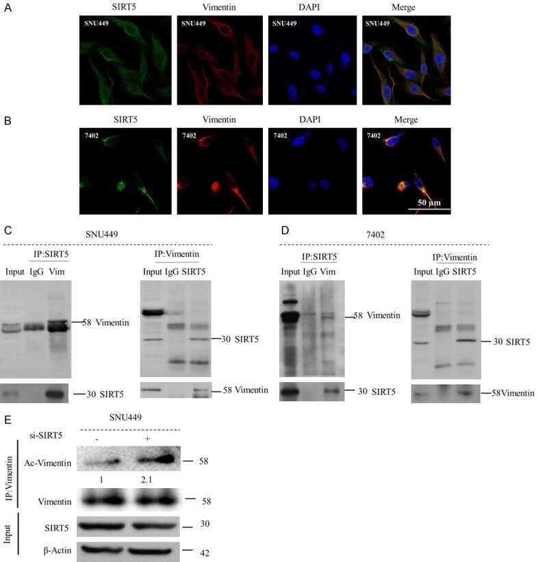
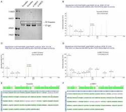
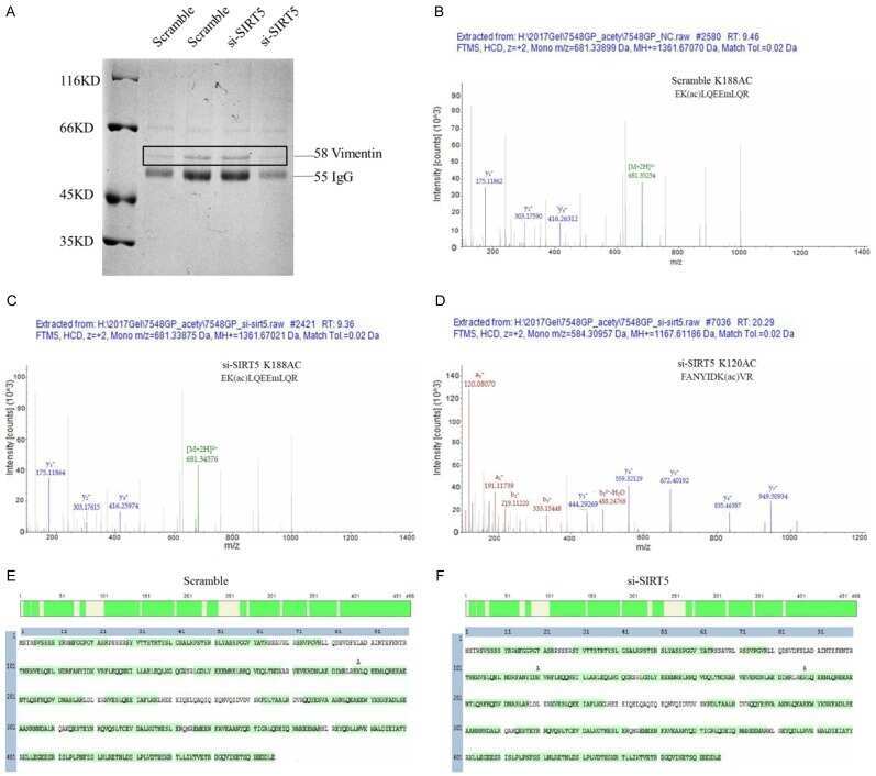
Vimentin acetylation is involved in SIRT5-mediated hepatocellular carcinoma migration.
Forkhead box C2 promotes the invasion ability of human trophoblast cells through Hedgehog (Hh) signaling pathway.
Kallikrein-related peptidase 4 induces cancer-associated fibroblast features in prostate-derived stromal cells.
Cytosolic Hsp90α and its mitochondrial isoform Trap1 are differentially required in a breast cancer model.
Real-Time Tracking of BODIPY-C12 Long-Chain Fatty Acid in Human Term Placenta Reveals Unique Lipid Dynamics in Cytotrophoblast Cells.
Doxorubicin and cisplatin induce apoptosis in ovarian stromal cells obtained from cryopreserved human ovarian tissue.
Cisplatin promotes mesenchymal-like characteristics in osteosarcoma through Snail.
Michalik S, Siegerist F, Palankar R, Franzke K, Schindler M, Reder A, Seifert U, Cammann C, Wesche J, Steil L, Hentschker C, Gesell-Salazar M, Reisinger E, Beer M, Endlich N, Greinacher A, Völker U
Haematologica 2022 Apr 1;107(4):947-957
Haematologica 2022 Apr 1;107(4):947-957
Identification of the Molecular Basis of Nanocurcumin-Induced Telocyte Preservation within the Colon of Ulcerative Colitis Rat Model.
Arafat EA, Marzouk RE, Mostafa SA, Hamed WHE
Mediators of inflammation 2021;2021:7534601
Mediators of inflammation 2021;2021:7534601
E2F5 Promotes the Malignancy of Ovarian Cancer Via the Regulation of Hippo and Wnt Pathways.
Malgundkar SH, Burney I, Al Moundhri M, Al Kalbani M, Lakhtakia R, Okamoto A, Tamimi Y
Genetic testing and molecular biomarkers 2021 Mar;25(3):179-186
Genetic testing and molecular biomarkers 2021 Mar;25(3):179-186
CD44 alternative splicing senses intragenic DNA methylation in tumors via direct and indirect mechanisms.
Batsché E, Yi J, Mauger O, Kornobis E, Hopkins B, Hanmer-Lloyd C, Muchardt C
Nucleic acids research 2021 Jun 21;49(11):6213-6237
Nucleic acids research 2021 Jun 21;49(11):6213-6237
D-Propranolol Impairs EGFR Trafficking and Destabilizes Mutant p53 Counteracting AKT Signaling and Tumor Malignancy.
Barra J, Cerda-Infante J, Sandoval L, Gajardo-Meneses P, Henriquez JF, Labarca M, Metz C, Venegas J, Retamal C, Oyanadel C, Cancino J, Soza A, Cuello MA, Roa JC, Montecinos VP, Gonzalez A
Cancers 2021 Jul 20;13(14)
Cancers 2021 Jul 20;13(14)
Establishment and Characterization of a Novel Dedifferentiated Chondrosarcoma Cell Line DDCS2.
Li X, Dean DC, Ferreira A, Nelson SD, Hornicek FJ, Yu S, Duan Z
Cancer control : journal of the Moffitt Cancer Center 2021 Jan-Dec;28:10732748211045274
Cancer control : journal of the Moffitt Cancer Center 2021 Jan-Dec;28:10732748211045274
Ginsenoside Rg3 inhibits osteosarcoma progression by reducing circ_0003074 expression in a miR-516b-5p/KPNA4-dependent manner.
Wang T, Zhang C, Wang S
Journal of orthopaedic surgery and research 2021 Dec 20;16(1):724
Journal of orthopaedic surgery and research 2021 Dec 20;16(1):724
Mitotic Activation Around Wound Edges and Epithelialization Repair in UVB-Induced Capsular Cataracts.
Wei Z, Hao C, Srinivasagan R, Wu H, Chen JK, Fan X
Investigative ophthalmology & visual science 2021 Dec 1;62(15):29
Investigative ophthalmology & visual science 2021 Dec 1;62(15):29
Differential expression and inhibitory effects of aquaporins on the development of adenomyosis.
Jia L, Liu Y, Han Y, Zhou X, Wang F
Molecular medicine reports 2020 Nov;22(5):3840-3850
Molecular medicine reports 2020 Nov;22(5):3840-3850
FAT4 silencing promotes epithelial-to-mesenchymal transition and invasion via regulation of YAP and β-catenin activity in ovarian cancer.
Malgundkar SH, Burney I, Al Moundhri M, Al Kalbani M, Lakhtakia R, Okamoto A, Tamimi Y
BMC cancer 2020 May 4;20(1):374
BMC cancer 2020 May 4;20(1):374
Periconceptional alcohol exposure causes female-specific perturbations to trophoblast differentiation and placental formation in the rat.
Kalisch-Smith JI, Steane SE, Simmons DG, Pantaleon M, Anderson ST, Akison LK, Wlodek ME, Moritz KM
Development (Cambridge, England) 2019 Jun 10;146(11)
Development (Cambridge, England) 2019 Jun 10;146(11)
Citrullination Controls Dendritic Cell Transdifferentiation into Osteoclasts.
Krishnamurthy A, Ytterberg AJ, Sun M, Sakuraba K, Steen J, Joshua V, Tarasova NK, Malmström V, Wähämaa H, Réthi B, Catrina AI
Journal of immunology (Baltimore, Md. : 1950) 2019 Jun 1;202(11):3143-3150
Journal of immunology (Baltimore, Md. : 1950) 2019 Jun 1;202(11):3143-3150
Vimentin acetylation is involved in SIRT5-mediated hepatocellular carcinoma migration.
Guo D, Song X, Guo T, Gu S, Chang X, Su T, Yang X, Liang B, Huang D
American journal of cancer research 2018;8(12):2453-2466
American journal of cancer research 2018;8(12):2453-2466
Forkhead box C2 promotes the invasion ability of human trophoblast cells through Hedgehog (Hh) signaling pathway.
Zhang Y, Zhang Y
Cell biology international 2018 Jul;42(7):859-866
Cell biology international 2018 Jul;42(7):859-866
Kallikrein-related peptidase 4 induces cancer-associated fibroblast features in prostate-derived stromal cells.
Kryza T, Silva LM, Bock N, Fuhrman-Luck RA, Stephens CR, Gao J, Samaratunga H, Australian Prostate Cancer BioResource, Lawrence MG, Hooper JD, Dong Y, Risbridger GP, Clements JA
Molecular oncology 2017 Oct;11(10):1307-1329
Molecular oncology 2017 Oct;11(10):1307-1329
Cytosolic Hsp90α and its mitochondrial isoform Trap1 are differentially required in a breast cancer model.
Vartholomaiou E, Madon-Simon M, Hagmann S, Mühlebach G, Wurst W, Floss T, Picard D
Oncotarget 2017 Mar 14;8(11):17428-17442
Oncotarget 2017 Mar 14;8(11):17428-17442
Real-Time Tracking of BODIPY-C12 Long-Chain Fatty Acid in Human Term Placenta Reveals Unique Lipid Dynamics in Cytotrophoblast Cells.
Kolahi K, Louey S, Varlamov O, Thornburg K
PloS one 2016;11(4):e0153522
PloS one 2016;11(4):e0153522
Doxorubicin and cisplatin induce apoptosis in ovarian stromal cells obtained from cryopreserved human ovarian tissue.
Fabbri R, Macciocca M, Vicenti R, Paradisi R, Klinger FG, Pasquinelli G, Spisni E, Seracchioli R, Papi A
Future oncology (London, England) 2016 Jul;12(14):1699-711
Future oncology (London, England) 2016 Jul;12(14):1699-711
Cisplatin promotes mesenchymal-like characteristics in osteosarcoma through Snail.
Fang S, Yu L, Mei H, Yang J, Gao T, Cheng A, Guo W, Xia K, Liu G
Oncology letters 2016 Dec;12(6):5007-5014
Oncology letters 2016 Dec;12(6):5007-5014
No comments: Submit comment
Supportive validation
- Submitted by
- Invitrogen Antibodies (provider)
- Main image
- Experimental details
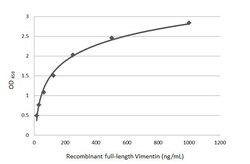
- Sandwich ELISA detection of recombinant full-length Vimentin protein using Vimentin Monoclonal Antibody (GT7812) (Product # MA5-31486) as capture antibody at concentration of 5 µg/mL and Vimentin Polyclonal Antibody (Product # PA5-27231) as detection antibody at concentration of 1 µg/mL. Rabbit IgG antibody (HRP) was diluted at 1:10,000 and used to detect the primary antibody.

- Submitted by
- Invitrogen Antibodies (provider)
- Main image

- Experimental details
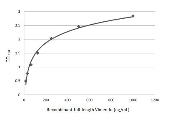
- Sandwich ELISA detection of recombinant full-length Vimentin protein using Vimentin Monoclonal Antibody (GT7812) (Product # MA5-31486) as capture antibody at concentration of 5 µg/mL and Vimentin Polyclonal Antibody (Product # PA5-27231) as detection antibody at concentration of 1 µg/mL. Rabbit IgG antibody (HRP) was diluted at 1:10,000 and used to detect the primary antibody.
- Submitted by
- Invitrogen Antibodies (provider)
- Main image
- Experimental details
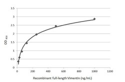
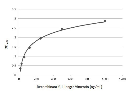
- Sandwich ELISA detection of recombinant full-length Vimentin protein using a Vimentin Monoclonal Antibody as capture antibody at concentration of 5 µg/mL and Vimentin Polyclonal Antibody (Product # PA5-27231) as detection antibody at concentration of 1 µg/mL. Rabbit IgG antibody (HRP) was diluted at 1:10,000 and used to detect the primary antibody.
Supportive validation
- Submitted by
- Invitrogen Antibodies (provider)
- Main image

- Experimental details
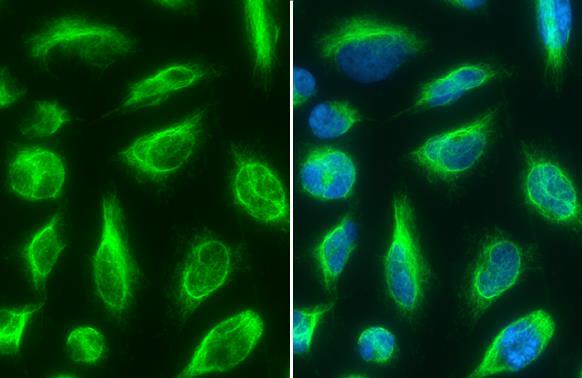
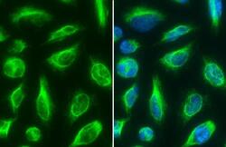
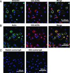
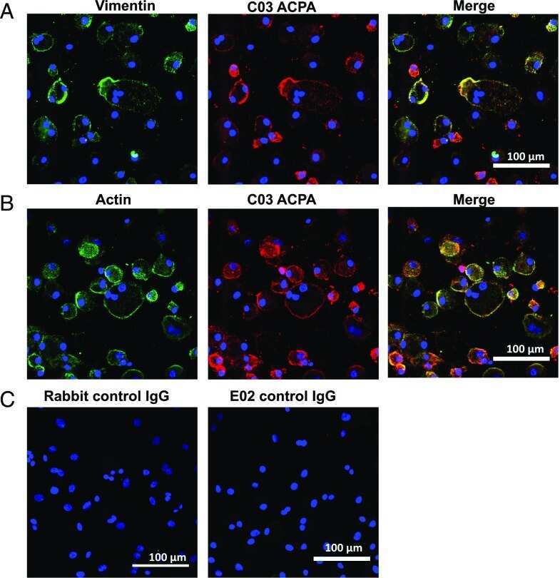
- Immunocytochemistry-Immunofluorescence analysis of Vimentin was performed in HeLa cells fixed in 4% paraformaldehyde at RT for 15 min. Green: Vimentin Polyclonal Antibody (Product # PA5-27231) diluted at 1:500. Blue: Hoechst 33342 staining. Scale bar = 10 µm.

- Submitted by
- Invitrogen Antibodies (provider)
- Main image

- Experimental details
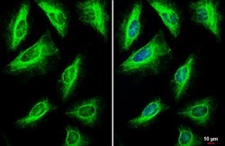
- Vimentin Polyclonal Antibody detects Vimentin protein at cytoskeleton by immunofluorescent analysis. Sample: HeLa cells were fixed in 4% paraformaldehyde at RT for 15 min. Green: Vimentin stained by Vimentin Polyclonal Antibody (Product # PA5-27231) diluted at 1:500. Blue: Fluoroshield with DAPI .
- Submitted by
- Invitrogen Antibodies (provider)
- Main image
- Experimental details
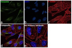
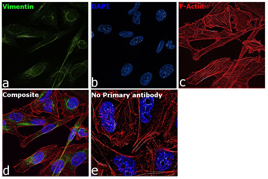
- Immunofluorescence analysis of Vimentin was performed using 70 confluent log phase MDA-MB-231 cells. The cells were fixed with 4% paraformaldehyde for 10 minutes, permeabilized with 0.1% Triton™ X-100 for 15 minutes, and blocked with 2% BSA for 45 minutes at room temperature. The cells were labeled with Vimentin Polyclonal Antibody (Product # PA5-27231) at 1:100 dilution in 0.1% BSA, incubated at 4 degree celsius overnight and then labeled with Donkey anti-Rabbit IgG (H+L) Highly Cross-Adsorbed Secondary Antibody, Alexa Fluor Plus 488 (Product # A32790), (1:2000 dilution), for 45 minutes at room temperature (Panel a: Green). Nuclei (Panel b:Blue) were stained with ProLong™ Diamond Antifade Mountant with DAPI (Product # P36962). F-actin (Panel c: Red) was stained with Rhodamine Phalloidin (Product # R415, 1:300 dilution). Panel d represents the merged image showing cytoskeleton localization. Panel e represents control cells with no primary antibody to assess background. The images were captured at 60X magnification.

- Submitted by
- Invitrogen Antibodies (provider)
- Main image
- Experimental details
- Vimentin Polyclonal Antibody detects Vimentin protein at cytoskeleton by immunofluorescent analysis. Sample: HeLa cells were fixed in 4% paraformaldehyde at RT for 15 min. Green: Vimentin stained by Vimentin Polyclonal Antibody (Product # PA5-27231) diluted at 1:500. Blue: Fluoroshield with DAPI .

- Submitted by
- Invitrogen Antibodies (provider)
- Main image
- Experimental details
- Immunocytochemistry-Immunofluorescence analysis of Vimentin was performed in HeLa cells fixed in 4% paraformaldehyde at RT for 15 min. Green: Vimentin Polyclonal Antibody (Product # PA5-27231) diluted at 1:500. Blue: Hoechst 33342 staining. Scale bar = 10 µm.

- Submitted by
- Invitrogen Antibodies (provider)
- Main image

- Experimental details
- Immunofluorescence analysis of Vimentin was performed using 70 confluent log phase MDA-MB-231 cells. The cells were fixed with 4% paraformaldehyde for 10 minutes, permeabilized with 0.1% Triton™ X-100 for 15 minutes, and blocked with 2% BSA for 45 minutes at room temperature. The cells were labeled with Vimentin Polyclonal Antibody (Product # PA5-27231) at 1:100 dilution in 0.1% BSA, incubated at 4 degree celsius overnight and then labeled with Donkey anti-Rabbit IgG (H+L) Highly Cross-Adsorbed Secondary Antibody, Alexa Fluor Plus 488 (Product # A32790), (1:2000 dilution), for 45 minutes at room temperature (Panel a: Green). Nuclei (Panel b:Blue) were stained with ProLong™ Diamond Antifade Mountant with DAPI (Product # P36962). F-actin (Panel c: Red) was stained with Rhodamine Phalloidin (Product # R415, 1:300 dilution). Panel d represents the merged image showing cytoskeleton localization. Panel e represents control cells with no primary antibody to assess background. The images were captured at 60X magnification.
Supportive validation
- Submitted by
- Invitrogen Antibodies (provider)
- Main image
- Experimental details
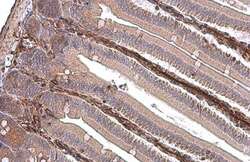
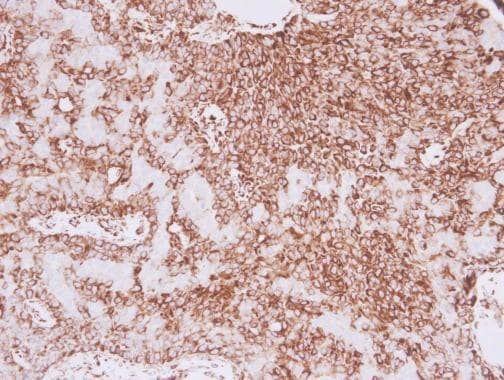
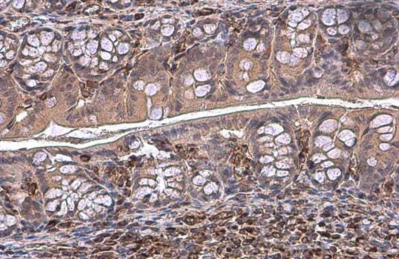
- Immunohistochemistry (Paraffin) analysis of Vimentin was performed in paraffin-embedded mouse intestine tissue using Vimentin Polyclonal Antibody (Product # PA5-27231) at a dilution of 1:500. Antigen Retrieval: Citrate buffer, pH 6.0, 15 min.
- Submitted by
- Invitrogen Antibodies (provider)
- Main image
- Experimental details
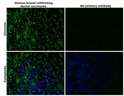
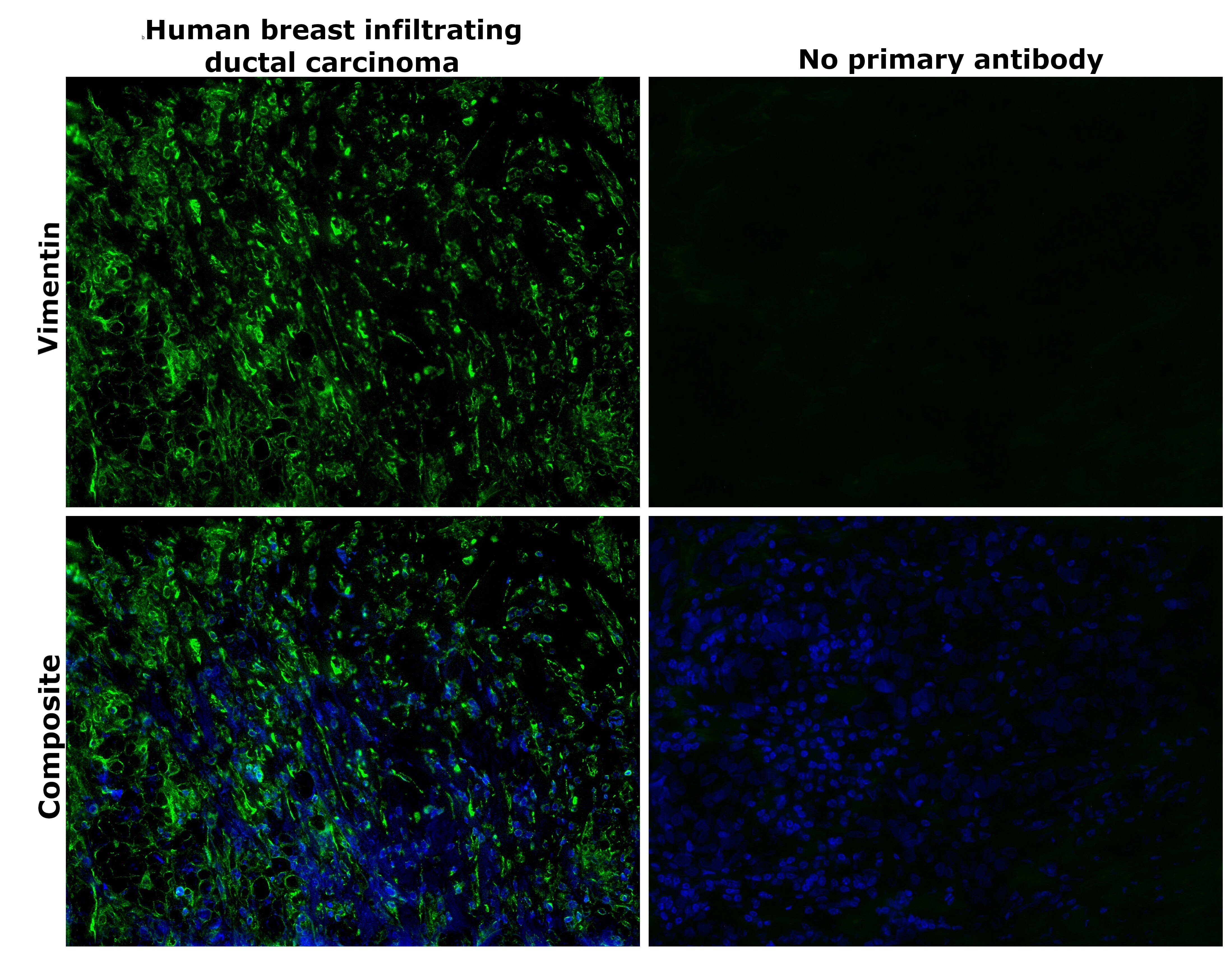
- Immunohistochemical analysis of Vimentin was performed using formalin-fixed paraffin-embedded human breast infiltrating ductal carcinoma tissue sections. To expose the target protein, heat-induced epitope retrieval was performed on de-paraffinized sections using eBioscience™ IHC Antigen Retrieval Solution - Low pH (10X) (Product # 00-4955-58) diluted to 1X solution in water in a decloaking chamber at 110 degree Celsius for 15 minutes. Following antigen retrieval, the sections were blocked with 2% normal goat serum in 1X PBS for 45 minutes at room temperature and then probed with or without Vimentin Polyclonal Antibody (Product # PA5-27231) at 1:100 dilution in 0.1% normal goat serum overnight at 4 degree Celsius in a humidified chamber. Detection was performed using Goat anti-Rabbit IgG (H+L) Highly Cross-Adsorbed Secondary Antibody, Alexa Fluor™ Plus 488 (Product # A32731) at a dilution of 1:2000 in 0.1% normal goat serum for 45 minutes at room temperature. Nuclei were stained with DAPI (Product # D1306) and the sections were mounted using ProLong™ Glass Antifade Mountant (Product # P36984). The images were captured on EVOS™ M7000 Imaging System (Product # AMF7000) at 20X magnification and externally deconvoluted.
- Submitted by
- Invitrogen Antibodies (provider)
- Main image
- Experimental details
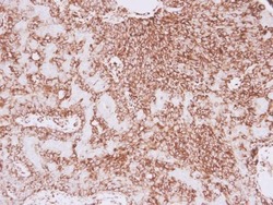
- Immunohistochemistry (Paraffin) analysis of Vimentin was performed in paraffin-embedded human lung adenocarcinoma tissue using Vimentin Polyclonal Antibody (Product # PA5-27231) at a dilution of 1:500.
- Submitted by
- Invitrogen Antibodies (provider)
- Main image

- Experimental details
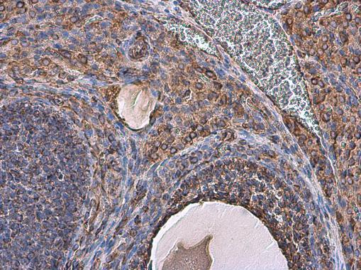
- Immunohistochemistry (Paraffin) analysis of Vimentin was performed in paraffin-embedded rat ovary tissue using Vimentin Polyclonal Antibody (Product # PA5-27231) at a dilution of 1:500.
- Submitted by
- Invitrogen Antibodies (provider)
- Main image
- Experimental details
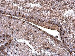
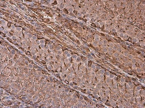
- Immunohistochemistry (Paraffin) analysis of Vimentin was performed in paraffin-embedded mouse testis tissue using Vimentin Polyclonal Antibody (Product # PA5-27231) at a dilution of 1:500.
- Submitted by
- Invitrogen Antibodies (provider)
- Main image
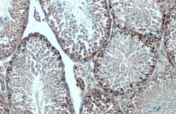
- Experimental details
- Vimentin Polyclonal Antibody detects Vimentin protein at cytoplasm by immunohistochemical analysis. Sample: Paraffin-embedded mouse testis. Vimentin stained by Vimentin Polyclonal Antibody (Product # PA5-27231) diluted at 1:500. Antigen Retrieval: Citrate buffer, pH 6.0, 15.
- Submitted by
- Invitrogen Antibodies (provider)
- Main image

- Experimental details
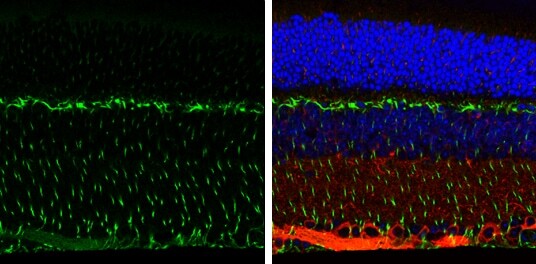
- Immunohistochemistry (Paraffin) analysis of Vimentin was performed in paraffin-Embedded adult mouse retina tissue using Green: Glutamine Synthetase Polyclonal Antibody (Product # PA5-28940) at a dilution of 1:250. Red: beta Tubulin 3/ TUJ1, stained by beta Tubulin 3/ TUJ1 antibody diluted at 1:500. Blue: Fluoroshield with DAPI.
- Submitted by
- Invitrogen Antibodies (provider)
- Main image
- Experimental details
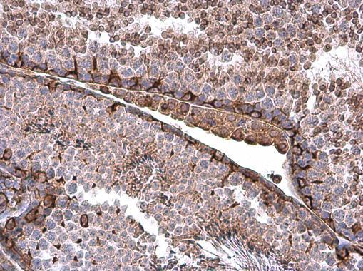
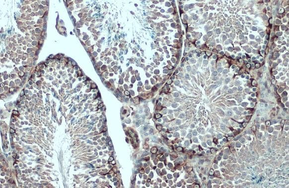
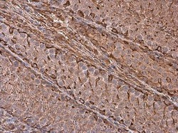
- Immunohistochemistry (Paraffin) analysis of Vimentin was performed in paraffin-embedded rat testis tissue using Vimentin Polyclonal Antibody (Product # PA5-27231) at a dilution of 1:500.
- Submitted by
- Invitrogen Antibodies (provider)
- Main image
- Experimental details
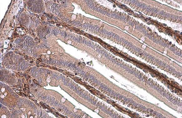
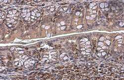
- Immunohistochemistry (Paraffin) analysis of Vimentin was performed in paraffin-embedded mouse colon tissue using Vimentin Polyclonal Antibody (Product # PA5-27231) at a dilution of 1:500. Antigen Retrieval: Citrate buffer, pH 6.0, 15 min.
Supportive validation
- Submitted by
- Invitrogen Antibodies (provider)
- Main image

- Experimental details
- NULL
- Submitted by
- Invitrogen Antibodies (provider)
- Main image

- Experimental details
- NULL
- Submitted by
- Invitrogen Antibodies (provider)
- Main image

- Experimental details
- NULL
- Submitted by
- Invitrogen Antibodies (provider)
- Main image
- Experimental details
- NULL
- Submitted by
- Invitrogen Antibodies (provider)
- Main image
- Experimental details
- NULL
- Submitted by
- Invitrogen Antibodies (provider)
- Main image
- Experimental details
- NULL
- Submitted by
- Invitrogen Antibodies (provider)
- Main image
- Experimental details
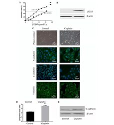
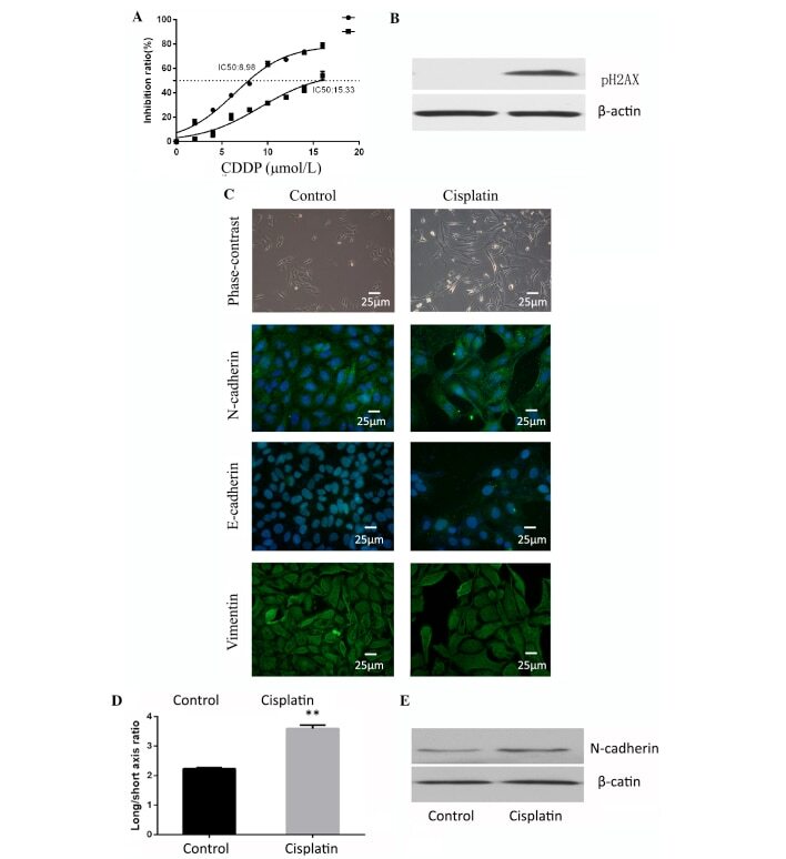
- Figure 1. Cisplatin induces epithelial-mesenchymal transition in osteosarcoma. (A) Cells treated with cisplatin were observed to have increased resistance to cisplatin. (B) Cells treated with cisplatin demonstrated high expression of pH2AX and confirmed the effectiveness of cisplatin. (C) Cell shape was observed by phase contrast microscopy and immunocytofluorescence. Staining of E-cadherin, N-cadherin and vimentin for the two groups of cells was observed by fluorescence microscope (magnification, x400; Scale, 25 um). Cells treated with cisplatin had higher N-cadherin expression. (D) Cisplatin-treated group cells had a higher average ratio of long/short axis. **P
- Submitted by
- Invitrogen Antibodies (provider)
- Main image
- Experimental details
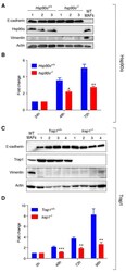
- Figure 4 hsp90alpha -/- and trap1 -/- cells of epithelial origin derived from mammary tumors show decreased proliferation A . and C . Immunoblots confirming epithelial origin of isolated cells, showing expression of E-cadherin and lack of expression of vimentin; cells were isolated from 3 Hsp90alpha +/+ and 3 hsp90alpha -/- mice, 2 Trap1 +/+ and 2 trap1 -/- animals (2 tumors per mouse); protein extracts from primary mouse adult fibroblasts (MAFs) were used as a positive control for vimentin expression; actin was used as loading control. B . hsp90alpha -/- cells exhibited a significant decrease in growth rates (measured by MTT assay) at 48 (* p < 0.05) and 72 hours (** p < 0.01) after seeding. D . A decrease in proliferation rates was observed in trap1 -/- cells (measured by standard cell counting) at 48 (*** p < 0.001), 72 (** p < 0.01) and 96 hours (** p < 0.01) post seeding; results of three independent experiments are illustrated as a fold change to the absorbance at 24 hours after seeding (Hsp90alpha) or to the number of cells seeded (Trap1).

- Submitted by
- Invitrogen Antibodies (provider)
- Main image

- Experimental details
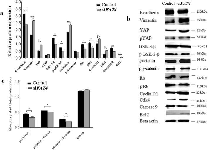
- Fig. 4 Regulatory effects of FAT4 on the expression of proteins involved in EMT, Hippo, Wnt-beta-catenin, apoptotic, and retinoblastoma pathways by Western blotting. a . Relative expression variation of proteins. The expression of Vimentin ( p = 0.0001), YAP ( p = 0.0018), beta-catenin ( p = 0.001), Bcl2 ( p = 0.0001), cyclin D1 ( p = 0.0017) and cdk4 ( p = 0.0025) was higher in FAT4 siRNA treated cells as compared to control. beta-actin was used as an internal control. b . Western blot demonstrating bands for protein expression in control and FAT4 knocked cells. Full-length blots are presented in Supplementary figure S 1 . c . The ratio of phosphorylated to total YAP, GSK-3beta, beta-catenin, and retinoblastoma proteins following FAT4 repression. The pYAP/ YAP ratio was lower in FAT4 siRNA treated cells as compared to the control ( p = 0.0286). Similarly, pGSK-3-beta/GSK-3-beta ratio, and pbeta-catenin/beta-catenin ratio was lower in FAT4 siRNA treated cells ( p = 0.018, and p = 0.001 respectively) as compared to control. There was no significant difference in pRb/Rb ratio between FAT4 siRNA treated cells and control. MCAS cells treated with scrambled siRNA was used as control. Data represent mean and standard deviation from at least three independent experiments performed in triplicates
- Submitted by
- Invitrogen Antibodies (provider)
- Main image
- Experimental details
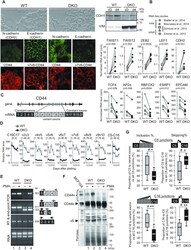
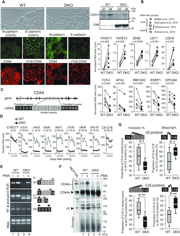
- Figure 1. Loss of DNA methylation leads to epithelial-to-mesenchymal Transition (EMT) and a decrease in variant CD44 protein isoforms. ( A ) Left panels: immunofluorescence analysis of WT or DKO HCT116 cells with mouse antibodies (red panels) against CD44, either with Hermes3 antibody (CD44) recognizing an epitope in the C5 exon () or with an antibody against v7v8 variant exons (v7v8-CD44), and analysis with rabbit antibodies (green panels) against E (CDH1) or N-(CDH2) cadherins. Right panels: western blot analysis of E-cadherin (CDH1) and Vimentin (VIM) expression levels in whole cell extracts from WT or DKO HCT116 cells, 3 and 4 days after plating as in Figure 2C which shows identical protein loading between samples. ( B ) RNA-seq from 4 studies were combined ( n = 5 samples) and matched for studies and types of extracts. The average of duplicates from the Blatter's study has been considered as one sample. Error bars show the mean absolute deviation (dev.) (see Supplementary Figure S1C ). The relative levels were cpm normalized for each librairy sizes. Statistical analysis has been carried out on Rlog (DESeq2) normalized counts using a paired t -test (two-tailed) to compare WT to DKO levels. Top and bottom panels show mesenchymal-specific and epithelial-specific genes, respectively. ( C ) Map of the human CD44 gene and mRNA. ( D ) Relative mRNA levels of CD44 variant exons in WT and DKO HCT116 cells at each indicated day after plating, analysed by RT-qPCR using primers pair
- Submitted by
- Invitrogen Antibodies (provider)
- Main image
- Experimental details
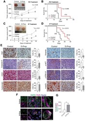
- Figure 4 D-Prop inhibits tumor growth and extends the survival of G-415 R282W xenografted NSG mice, with congruent immunohistochemical changes. Treatment started when G-415 R282W xenografts reached 50 mm 3 and consisted on single daily doses of D-Prop administered for 15 days, either per oral (PO) (70 mg/kg/day) ( A , B ) or intraperitoneal (IP) (20 mg/kg/day) ( C , D ), as indicated. ( A , C ) Tumor volume curves reflect decreased tumor growth under both D-Prop treatments, either PO (* p = 0.05, n = 5 mice per group) or IP (** p = 0.01; n = 10 mice per group). Insets show representative images of tumors. ( B , D ) Kaplan-Meier analysis shows extended survival under both D-Prop treatments, PO ( B ) (median survival = 47 versus 28 days; p = 0.008 log-rank test; n = 5) and IP ( D ) (median survivals = 28.5 versus 20 days; p = 0.013, log-rank test; n = 10). ( E ) Immunohistochemistry (IHC) of tumors at the end of IP D-Prop treatment. IHC images (left and right panels) and graphs with the percentage of cells stained for the indicated proteins show decreased levels of the proliferation marker Ki67, endothelial marker CD31, mesenchymal marker vimentin, signaling proteins pAKT and pERK and mutp53, whereas the apoptotic marker cleavage caspase-3 increased. Cytokeratin show no changes. Graphs shows means +- SEM (* p < 0.05; ** p < 0.01; t -test). ( F ) D-Prop induces EGFR internalization in G-415 R282W xenografts. Immunofluorescence of EGFR (green) with TfnR (magenta) in cryostat sect

- Submitted by
- Invitrogen Antibodies (provider)
- Main image
- Experimental details
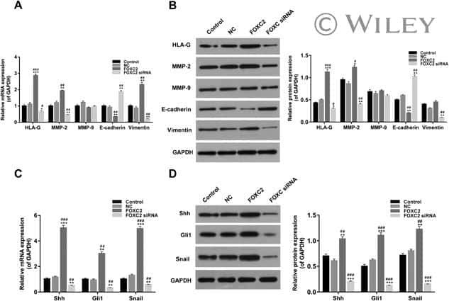
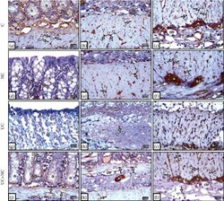
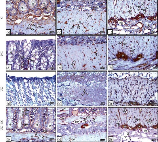
- Figure 7 Photomicrographs of vimentin immunohistochemical-stained sections in the colon. (a-c) The control group and (d-f) NC group both show plentiful vimentin-positive TC cells (arrow) with long Tps (arrowhead) close to the crypt epithelium, muscularis mucosa, submucosa, musculosa, myenteric plexus, and around blood vessel (BV). (g-i) The UC group reveals hardly seen TCs in the mucosa and submucosa and few numbers of cells in the musculosa and myenteric plexus. (j-l) The UC+NC group shows abundant TCs in the mucosa, submucosa, musculosa, and myenteric plexus (*) (vimentin immunostaining X400 insert in k X1000).
- Submitted by
- Invitrogen Antibodies (provider)
- Main image

- Experimental details
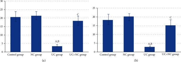
- Figure 8 (a) Analysis of the number of CD 34 positive immune-stained TC/HPF in the study groups (6 rats/group). A: significant difference in relation to control group. B: significant difference in relation to NC group. C: significant difference in relation to UC group. (b) Analysis of the vimentin-positive immune-stained cells TC/HPF in the study groups (6 rats/group). A: significant difference in relation to control group. B: significant difference in relation to NC group. C: significant difference in relation to UC group.
- Submitted by
- Invitrogen Antibodies (provider)
- Main image
- Experimental details
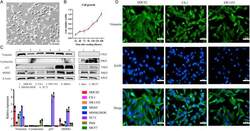
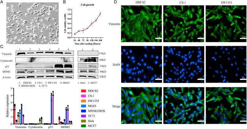
- Figure 2. Morphologic characteristics and antigenic marker expression of the novel DDCS2 cell line. (A) DDCS2 cells display spindle to polygonal shape in vitro under light microscopy (200x, scale bar: 50 mum). (B) The growth curves of DDCS2 were plotted using cells plated in 96-well plates. (C) Western blots revealed the expression of vimentin, cytokeratin, p53, and MDM2 in various sarcoma cell lines compared with two epithelial carcinoma cell lines. Relative expressions of vimentin, cytokeratin, p53, and MDM2 were figured in different sarcoma and epithelial carcinoma cell lines compared with beta-actin. (D) Immunofluorescence analysis of vimentin was conducted in DDCS2, CS-1, and SW1353 cell lines (200x, scale bar: 50 mum).
- Submitted by
- Invitrogen Antibodies (provider)
- Main image
- Experimental details
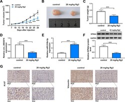
- Fig. 8 Rg3 treatment inhibited OS cell malignancy in vivo. A - C The effect of Rg3 treatment on tumorigenesis of MG63 cells. D - F The impacts of Rg3 treatment on the expression of circ_0003074, miR-516b-5p and KPNA4 were determined by qRT-PCR or Western blotting in the primary tumors from MG63 cells. G The positive expression rate of Ki-67 was checked by IHC assay in the primary tumors from MG63 cells. ** P < 0.01 and *** P < 0.001
