Explore
 Validate
 Learn
Learn Western blot
 Immunocytochemistry
Antibody data
- Antibody Data
- Antigen structure
- References [19]
- Comments [0]
- Validations
- Immunocytochemistry [1]
- Immunohistochemistry [1]
Submit
Validation data
Reference
Comment
Report error
- Product number
- HPA001762 - Provider product page

- Provider
- Atlas Antibodies
- Proper citation
- Atlas Antibodies Cat#HPA001762, RRID:AB_1080568
- Product name
- Anti-VIM
- Antibody type
- Polyclonal
- Description
- Polyclonal Antibody against Human VIM, Gene description: vimentin, Validated applications: IHC, WB, ICC, Uniprot ID: P08670, Storage: Store at +4°C for short term storage. Long time storage is recommended at -20°C.
- Reactivity
- Human, Mouse
- Host
- Rabbit
- Conjugate
- Unconjugated
- Isotype
- IgG
- Vial size
- 100 µl
- Concentration
- 0.2 mg/ml
- Storage
- Store at +4°C for short term storage. Long time storage is recommended at -20°C.
- Handling
- The antibody solution should be gently mixed before use.
Submitted references Silencing of GRHL2 induces epithelial‑to‑mesenchymal transition in lung cancer cell lines with different effects on proliferation and clonogenic growth
TGFß1 Stimulates Lymphatic Endothelial Cells to Produce IL7 and IL15, Which Act as Chemotactic Factors for Breast Cancer Cells with Mesenchymal Properties.
Neutrophil extracellular traps promote gastric cancer metastasis by inducing epithelial‑mesenchymal transition
Photothermal Therapy as Adjuvant to Surgery in an Orthotopic Mouse Model of Human Fibrosarcoma
In vitro and in vivo characterization of a recombinant rhesus cytomegalovirus containing a complete genome
Ginkgolic Acid, a SUMO-1 Inhibitor, Inhibits the Progression of Oral Squamous Cell Carcinoma by Alleviating SUMOylation of SMAD4
Systematic analysis reveals a functional role for STAMBPL1 in the epithelial-mesenchymal transition process across multiple carcinomas.
Development of a predictive model for stromal content in prostate cancer samples to improve signature performance
PEP06 polypeptide 30 exerts antitumour effect in colorectal carcinoma via inhibiting epithelial–mesenchymal transition
2-Arachidonoylglycerol Reduces Proteoglycans and Enhances Remyelination in a Progressive Model of Demyelination
A 3D in vitro model of the human breast duct: a method to unravel myoepithelial-luminal interactions in the progression of breast cancer.
Extracellular Vesicles from Metastatic Rat Prostate Tumors Prime the Normal Prostate Tissue to Facilitate Tumor Growth
Inhibition of Lysyl Oxidase and Lysyl Oxidase-Like Enzymes Has Tumour-Promoting and Tumour-Suppressing Roles in Experimental Prostate Cancer
Establishment of 3D Co-Culture Models from Different Stages of Human Tongue Tumorigenesis: Utility in Understanding Neoplastic Progression
Regulation of Glutamine Carrier Proteins by RNF5 Determines Breast Cancer Response to ER Stress-Inducing Chemotherapies
Changes in Cannabinoid Receptors, Aquaporin 4 and Vimentin Expression after Traumatic Brain Injury in Adolescent Male Mice. Association with Edema and Neurological Deficit
Systematic validation of antibody binding and protein subcellular localization using siRNA and confocal microscopy
Tumor suppressive microRNA-138 contributes to cell migration and invasion through its targeting of vimentin in renal cell carcinoma
Kawabe N, Matsuoka K, Komeda K, Muraki N, Takaba M, Togami Y, Ito Y, Yamada M, Sunaga N, Girard L, Minna J, Cai L, Xie Y, Tanaka I, Morise M, Sato M
Oncology Letters 2023;26(3)
Oncology Letters 2023;26(3)
TGFß1 Stimulates Lymphatic Endothelial Cells to Produce IL7 and IL15, Which Act as Chemotactic Factors for Breast Cancer Cells with Mesenchymal Properties.
Giotopoulou N, Shi W, Parniewska MM, Sun W, Fuxe J
Journal of mammary gland biology and neoplasia 2023 Dec 6;28(1):25
Journal of mammary gland biology and neoplasia 2023 Dec 6;28(1):25
Neutrophil extracellular traps promote gastric cancer metastasis by inducing epithelial‑mesenchymal transition
Zhu T, Zou X, Yang C, Li L, Wang B, Li R, Li H, Xu Z, Huang D, Wu Q
International Journal of Molecular Medicine 2021;48(1)
International Journal of Molecular Medicine 2021;48(1)
Photothermal Therapy as Adjuvant to Surgery in an Orthotopic Mouse Model of Human Fibrosarcoma
Simón M, Jørgensen J, Melander F, Andresen T, Christensen A, Kjaer A
Cancers 2021;13(22):5820
Cancers 2021;13(22):5820
FOXK1, Regulated by miR-365-3p, Promotes Cell Growth and EMT Indicates Unfavorable Prognosis in Breast Cancer
Gao F, Tian J
OncoTargets and Therapy 2020;Volume 13
OncoTargets and Therapy 2020;Volume 13
In vitro and in vivo characterization of a recombinant rhesus cytomegalovirus containing a complete genome
Kalejta R, Taher H, Mahyari E, Kreklywich C, Uebelhoer L, McArdle M, Moström M, Bhusari A, Nekorchuk M, E X, Whitmer T, Scheef E, Sprehe L, Roberts D, Hughes C, Jackson K, Selseth A, Ventura A, Cleveland-Rubeor H, Yue Y, Schmidt K, Shao J, Edlefsen P, Smedley J, Kowalik T, Stanton R, Axthelm M, Estes J, Hansen S, Kaur A, Barry P, Bimber B, Picker L, Streblow D, Früh K, Malouli D
PLOS Pathogens 2020;16(11):e1008666
PLOS Pathogens 2020;16(11):e1008666
Ginkgolic Acid, a SUMO-1 Inhibitor, Inhibits the Progression of Oral Squamous Cell Carcinoma by Alleviating SUMOylation of SMAD4
Liu K, Wang X, Li D, Xu D, Li D, Lv Z, Zhao D, Chu W, Wang X
Molecular Therapy - Oncolytics 2020;16
Molecular Therapy - Oncolytics 2020;16
Systematic analysis reveals a functional role for STAMBPL1 in the epithelial-mesenchymal transition process across multiple carcinomas.
Ambroise G, Yu TT, Zhang B, Kacal M, Hao Y, Queiroz AL, Ouchida AT, Lindskog C, Norberg E, Vakifahmetoglu-Norberg H
British journal of cancer 2020 Sep;123(7):1164-1177
British journal of cancer 2020 Sep;123(7):1164-1177
Development of a predictive model for stromal content in prostate cancer samples to improve signature performance
Boufaied N, Takhar M, Nash C, Erho N, Bismar T, Davicioni E, Thomson A
The Journal of Pathology 2019;249(4):411-424
The Journal of Pathology 2019;249(4):411-424
PEP06 polypeptide 30 exerts antitumour effect in colorectal carcinoma via inhibiting epithelial–mesenchymal transition
Yu S, Li L, Tian W, Nie D, Mu W, Qiu F, Liu Y, Liu X, Wang X, Du Z, Chu W, Yang B
British Journal of Pharmacology 2018;175(15):3111-3130
British Journal of Pharmacology 2018;175(15):3111-3130
2-Arachidonoylglycerol Reduces Proteoglycans and Enhances Remyelination in a Progressive Model of Demyelination
Feliú A, Bonilla del Río I, Carrillo-Salinas F, Hernández-Torres G, Mestre L, Puente N, Ortega-Gutiérrez S, López-Rodríguez M, Grandes P, Mecha M, Guaza C
The Journal of Neuroscience 2017;37(35):8385-8398
The Journal of Neuroscience 2017;37(35):8385-8398
A 3D in vitro model of the human breast duct: a method to unravel myoepithelial-luminal interactions in the progression of breast cancer.
Carter EP, Gopsill JA, Gomm JJ, Jones JL, Grose RP
Breast cancer research : BCR 2017 Apr 21;19(1):50
Breast cancer research : BCR 2017 Apr 21;19(1):50
Extracellular Vesicles from Metastatic Rat Prostate Tumors Prime the Normal Prostate Tissue to Facilitate Tumor Growth
Halin Bergström S, Hägglöf C, Thysell E, Bergh A, Wikström P, Lundholm M
Scientific Reports 2016;6(1)
Scientific Reports 2016;6(1)
Inhibition of Lysyl Oxidase and Lysyl Oxidase-Like Enzymes Has Tumour-Promoting and Tumour-Suppressing Roles in Experimental Prostate Cancer
Nilsson M, Adamo H, Bergh A, Halin Bergström S
Scientific Reports 2016;6(1)
Scientific Reports 2016;6(1)
Establishment of 3D Co-Culture Models from Different Stages of Human Tongue Tumorigenesis: Utility in Understanding Neoplastic Progression
Murdoch C, Sawant S, Dongre H, Singh A, Joshi S, Costea D, Mahadik S, Ahire C, Makani V, Dange P, Sharma S, Chaukar D, Vaidya M
PLOS ONE 2016;11(8):e0160615
PLOS ONE 2016;11(8):e0160615
Regulation of Glutamine Carrier Proteins by RNF5 Determines Breast Cancer Response to ER Stress-Inducing Chemotherapies
Jeon Y, Khelifa S, Ratnikov B, Scott D, Feng Y, Parisi F, Ruller C, Lau E, Kim H, Brill L, Jiang T, Rimm D, Cardiff R, Mills G, Smith J, Osterman A, Kluger Y, Ronai Z
Cancer Cell 2015;27(3):354-369
Cancer Cell 2015;27(3):354-369
Changes in Cannabinoid Receptors, Aquaporin 4 and Vimentin Expression after Traumatic Brain Injury in Adolescent Male Mice. Association with Edema and Neurological Deficit
Dehghani F, Lopez-Rodriguez A, Acaz-Fonseca E, Viveros M, Garcia-Segura L
PLOS ONE 2015;10(6):e0128782
PLOS ONE 2015;10(6):e0128782
Systematic validation of antibody binding and protein subcellular localization using siRNA and confocal microscopy
Stadler C, Hjelmare M, Neumann B, Jonasson K, Pepperkok R, Uhlén M, Lundberg E
Journal of Proteomics 2012;75(7):2236-2251
Journal of Proteomics 2012;75(7):2236-2251
Tumor suppressive microRNA-138 contributes to cell migration and invasion through its targeting of vimentin in renal cell carcinoma
YAMASAKI T, SEKI N, YAMADA Y, YOSHINO H, HIDAKA H, CHIYOMARU T, NOHATA N, KINOSHITA T, NAKAGAWA M, ENOKIDA H
International Journal of Oncology 2012;41(3):805-817
International Journal of Oncology 2012;41(3):805-817
No comments: Submit comment
Supportive validation
- Submitted by
- Atlas Antibodies (provider)
- Main image
- Experimental details
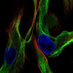
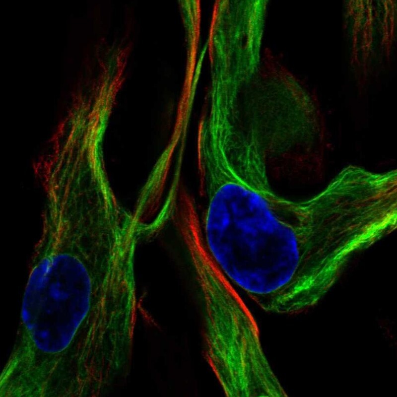
- Immunofluorescent staining of human cell line ASC TERT1 shows localization to intermediate filaments.
- Sample type
- Human
Supportive validation
- Submitted by
- Atlas Antibodies (provider)
- Enhanced method
- Orthogonal validation
- Main image
- Experimental details
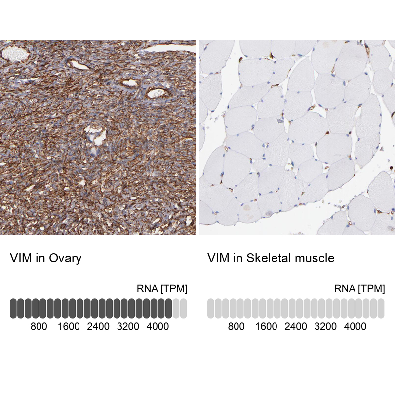
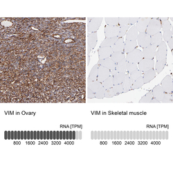
- Immunohistochemistry analysis in human ovary and skeletal muscle tissues using HPA001762 antibody. Corresponding VIM RNA-seq data are presented for the same tissues.
- Sample type
- Human
- Protocol
- Protocol