Explore
 Validate
 Learn
Learn Western blot
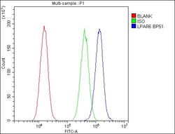
 Flow cytometry
Antibody data
- Antibody Data
- Antigen structure
- References [1]
- Comments [0]
- Validations
- Western blot [1]
Submit
Validation data
Reference
Comment
Report error
- Product number
- A05725-1 - Provider product page

- Provider
- Boster Biological Technology
- Product name
- Anti-P2RY5/LPAR6 Antibody Picoband™
- Antibody type
- Polyclonal
- Description
- Polyclonal antibody for P2Y5/LPAR6 detection. Host: Rabbit.Size: 100μg/vial. Tested applications: WB. Reactive species: Human. P2Y5/LPAR6 information: Subcellular Localization: Cell membrane; Tissue Specificity: Expressed ubiquitously, including in skin and hair follicle cells. Detected in both Henle's and Huxley's layers of the inner root sheath of the hair follicle and in suprabasal layers of the epidermis (at protein level). Expressed at low levels in peripheral blood leukocytes.
- Reactivity
- Human, Mouse, Rat
- Host
- Rabbit
- Vial size
- 100μg/vial
- Concentration
- 0.5-1mg/ml, actual concentration vary by lot. Use suggested dilution ratio to decide dilution procedure.
- Storage
- At -20°C for one year. After reconstitution, at 4°C for one month. It can also be aliquoted and stored frozen at -20°C for a longer time. Avoid repeated freezing and thawing.
- Handling
- Add 0.2ml of distilled water will yield a concentration of 500ug/ml.
Submitted references Electrochemically derived nanographene oxide activates endothelial tip cells and promotes angiogenesis by binding endogenous lysophosphatidic acid.
Liu W, Luo H, Wei Q, Liu J, Wu J, Zhang Y, Chen L, Ren W, Shao L
Bioactive materials 2022 Mar;9:92-104
Bioactive materials 2022 Mar;9:92-104
No comments: Submit comment
Supportive validation
- Submitted by
- Boster Biological Technology (provider)
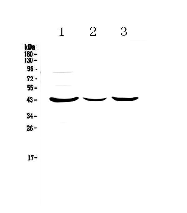
- Main image
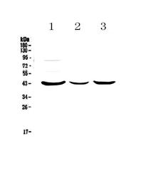
- Experimental details
- Western blot analysis of P2RY5 using anti-P2RY5 antibody (A05725-1). Electrophoresis was performed on a 5-20% SDS-PAGE gel at 70V (Stacking gel) / 90V (Resolving gel) for 2-3 hours. The sample well of each lane was loaded with 50ug of sample under reducing conditions. Lane 1: human MCF-7 whole cell lysates,Lane 2: human HepG2 whole cell lysates,Lane 3: human SK-OV-3 whole cell lysates. After Electrophoresis, proteins were transferred to a Nitrocellulose membrane at 150mA for 50-90 minutes. Blocked the membrane with 5% Non-fat Milk/ TBS for 1.5 hour at RT. The membrane was incubated with rabbit anti-P2RY5 antigen affinity purified polyclonal antibody (Catalog # A05725-1) at 0.5 μg/mL overnight at 4°C, then washed with TBS-0.1%Tween 3 times with 5 minutes each and probed with a goat anti-rabbit IgG-HRP secondary antibody at a dilution of 1:10000 for 1.5 hour at RT. The signal is developed using an Enhanced Chemiluminescent detection (ECL) kit (Catalog # EK1002) with Tanon 5200 system. A specific band was detected for P2RY5 at approximately 43KD. The expected band size for P2RY5 is at 39KD.
- Additional image