Explore
 Validate
 Learn
Learn Western blot
 Immunoprecipitation
Antibody data
- Antibody Data
- Antigen structure
- References [0]
- Comments [0]
- Validations
- Western blot [1]
- Immunocytochemistry [3]
Submit
Validation data
Reference
Comment
Report error
- Product number
- GTX19350 - Provider product page

- Provider
- GeneTex
- Proper citation
- GeneTex Cat#GTX19350, RRID:AB_423539
- Product name
- P-Cadherin antibody [6A9]
- Antibody type
- Monoclonal
- Reactivity
- Human, Mouse
- Host
- Mouse
No comments: Submit comment
Supportive validation
- Submitted by
- GeneTex (provider)
- Main image
- Experimental details
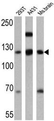
- WB analysis of 293T, A431 and mouse brain lysates (25 ug per lane) using P-Cadherin antibody [6A9] at a dilution of 1:500.
Supportive validation
- Submitted by
- GeneTex (provider)
- Main image
- Experimental details
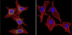
- Immunofluorescent analysis of P-Cadherin in A2058 Cells. P-Cadherin staining (green), F-Actin staining with Phalloidin (red) and nuclei with DAPI (blue) is shown. Cells were grown on slides and fixed with formaldehyde prior to staining. Cells were probed without (control) or with P-Cadherin antibody [6A9] at a dilution of 1:20 over night at 4 ¢XC, washed with PBS and incubated with a proper secondary antibody. Images were taken at 60X magnification.
- Submitted by
- GeneTex (provider)
- Main image

- Experimental details
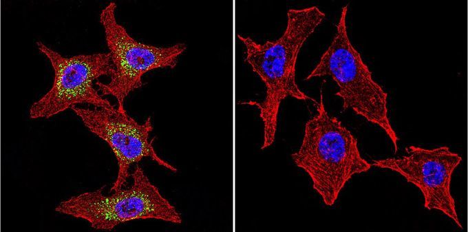
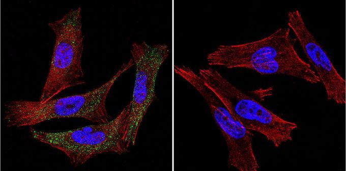
- ICC/IF analysis of BEAS-2B cells with (left) or without (right) P-Cadherin antibody [6A9] at a dilution of 1:20 (green). F-Actin staining with Phalloidin (red) and nuclei with DAPI (blue) is shown.
- Submitted by
- GeneTex (provider)
- Main image
- Experimental details
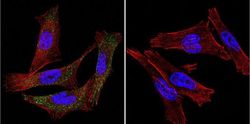
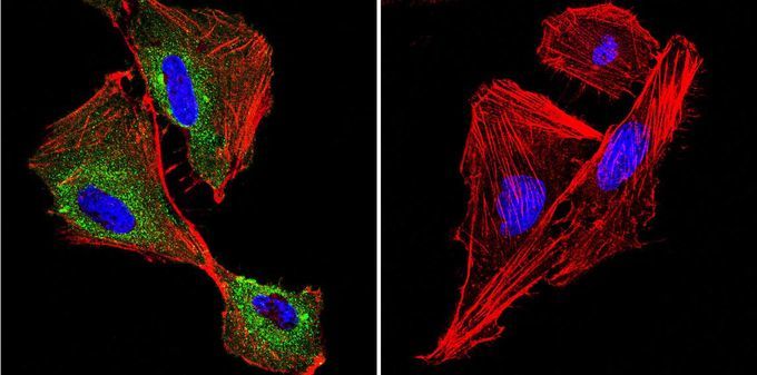
- ICC/IF analysis of HeLa cells with (left) or without (right) P-Cadherin antibody [6A9] at a dilution of 1:20 (green). F-Actin staining with Phalloidin (red) and nuclei with DAPI (blue) is shown.