Explore
 Validate
 Learn
Learn Western blot
 ELISA
ELISAAntibody data
- Antibody Data
- Antigen structure
- References [4]
- Comments [0]
- Validations
- Western blot [2]
- Immunocytochemistry [1]
- Immunohistochemistry [1]
Submit
Validation data
Reference
Comment
Report error
- Product number
- AF761 - Provider product page

- Provider
- R&D Systems
- Product name
- Mouse P-Cadherin Antibody
- Antibody type
- Polyclonal
- Description
- Antigen Affinity-purified. Detects P-Cadherin in ELISAs and Western blots. In sandwich immunoassays, less than 2% cross-reactivity with recombinant human (rh) P-Cadherin is observed and less than 0.3% cross-reactivity with recombinant mouse E-Cadherin, rhN-Cadherin, and rhCadherin-8 is observed.
- Reactivity
- Mouse
- Host
- Goat
- Conjugate
- Unconjugated
- Antigen sequence
Q8BSL6- Isotype
- IgG
- Vial size
- 100 ug
- Concentration
- LYOPH
- Storage
- Use a manual defrost freezer and avoid repeated freeze-thaw cycles. 12 months from date of receipt, -20 to -70 °C as supplied. 1 month, 2 to 8 °C under sterile conditions after reconstitution. 6 months, -20 to -70 °C under sterile conditions after reconstitution.
Submitted references Cadherins in the retinal pigment epithelium (RPE) revisited: P-cadherin is the highly dominant cadherin expressed in human and mouse RPE in vivo.
Stem Cell Lineage Infidelity Drives Wound Repair and Cancer.
Stem cell plasticity enables hair regeneration following Lgr5+ cell loss.
Role of cell and matrix-bound VEGF isoforms in lens development.
Yang X, Chung JY, Rai U, Esumi N
PloS one 2018;13(1):e0191279
PloS one 2018;13(1):e0191279
Stem Cell Lineage Infidelity Drives Wound Repair and Cancer.
Ge Y, Gomez NC, Adam RC, Nikolova M, Yang H, Verma A, Lu CP, Polak L, Yuan S, Elemento O, Fuchs E
Cell 2017 May 4;169(4):636-650.e14
Cell 2017 May 4;169(4):636-650.e14
Stem cell plasticity enables hair regeneration following Lgr5+ cell loss.
Hoeck JD, Biehs B, Kurtova AV, Kljavin NM, de Sousa E Melo F, Alicke B, Koeppen H, Modrusan Z, Piskol R, de Sauvage FJ
Nature cell biology 2017 Jun;19(6):666-676
Nature cell biology 2017 Jun;19(6):666-676
Role of cell and matrix-bound VEGF isoforms in lens development.
Saint-Geniez M, Kurihara T, D'Amore PA
Investigative ophthalmology & visual science 2009 Jan;50(1):311-21
Investigative ophthalmology & visual science 2009 Jan;50(1):311-21
No comments: Submit comment
Supportive validation
- Submitted by
- R&D Systems (provider)
- Main image
- Experimental details
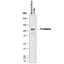
- Detection of P-Cadherin by Western Blot. Western blot shows lysates of mouse embryo tissue. PVDF membrane was probed with 0.5 µg/mL of Goat Anti-Mouse P-Cadherin Antigen Affinity-purified Polyclonal Antibody (Catalog # AF761) followed by HRP-conjugated Anti-Goat IgG Secondary Antibody (Catalog # HAF017). A specific band was detected for P-Cadherin at approximately 115 kDa (as indicated). This experiment was conducted under reducing conditions and using Immunoblot Buffer Group 1.

- Submitted by
- R&D Systems (provider)
- Main image
- Experimental details
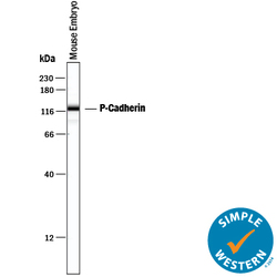
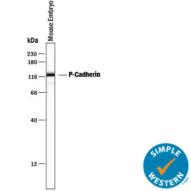
- Detection of Mouse P-Cadherin by Simple WesternTM. Simple Western lane view shows lysates of mouse embryo tissue, loaded at 0.2 mg/mL. A specific band was detected for P-Cadherin at approximately 115 kDa (as indicated) using 25 µg/mL of Goat Anti-Mouse P-Cadherin Antigen Affinity-purified Polyclonal Antibody (Catalog # AF761) followed by 1:50 dilution of HRP-conjugated Anti-Goat IgG Secondary Antibody (Catalog # HAF109). This experiment was conducted under reducing conditions and using the 12-230 kDa separation system.
Supportive validation
- Submitted by
- R&D Systems (provider)
- Main image
- Experimental details
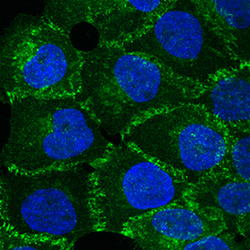
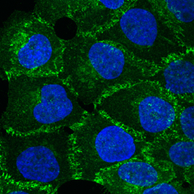
- P-Cadherin in A431 Human Cell Line. P-Cadherin was detected in immersion fixed A431 human epithelial carcinoma cell line using Goat Anti-Mouse P-Cadherin Antigen Affinity-purified Polyclonal Antibody (Catalog # AF761) at 10 µg/mL for 3 hours at room temperature. Cells were stained using the NorthernLights™ 493-conjugated Anti-Goat IgG Secondary Antibody (green; Catalog # NL003) and counterstained with DAPI (blue). Specific staining was localized to intercellular junctions. View our protocol for Fluorescent ICC Staining of Cells on Coverslips.
Supportive validation
- Submitted by
- R&D Systems (provider)
- Main image
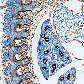
- Experimental details
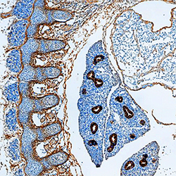
- P-Cadherin in Mouse Embryo. P-Cadherin was detected in immersion fixed frozen sections of mouse embryo (15 dpc) using Goat Anti-Mouse P-Cadherin Antigen Affinity-purified Polyclonal Antibody (Catalog # AF761) at 15 µg/mL overnight at 4 °C. Tissue was stained using the Anti-Goat HRP-DAB Cell & Tissue Staining Kit (brown; Catalog # CTS008) and counterstained with hematoxylin (blue). Specific staining was localized to connective tissue and lungs. View our protocol for Chromogenic IHC Staining of Frozen Tissue Sections.