Explore
 Validate
 Learn
Learn Western blot
 ELISA
ELISAAntibody data
- Antibody Data
- Antigen structure
- References [2]
- Comments [0]
- Validations
- Western blot [3]
- Immunocytochemistry [2]
- Flow cytometry [2]
Submit
Validation data
Reference
Comment
Report error
- Product number
- MAB861-100 - Provider product page

- Provider
- R&D Systems
- Product name
- Human P-Cadherin Antibody
- Antibody type
- Monoclonal
- Description
- Protein A or G purified from hybridoma culture supernatant. Detects human P-Cadherin in direct ELISAs and Western blots. In Western blots, does not cross-react with recombinant human (rh) Cadherin-8, recombinant mouse P-Cadherin, or rhVE-Cadherin.
- Reactivity
- Human
- Host
- Mouse
- Conjugate
- Unconjugated
- Antigen sequence
CAA45177- Isotype
- IgG
- Antibody clone number
- 104805
- Vial size
- 100 ug
- Storage
- Use a manual defrost freezer and avoid repeated freeze-thaw cycles. 12 months from date of receipt, -20 to -70 °C as supplied. 1 month, 2 to 8 °C under sterile conditions after reconstitution. 6 months, -20 to -70 °C under sterile conditions after reconstitution.
Submitted references Cadherin-23 mediates heterotypic cell-cell adhesion between breast cancer epithelial cells and fibroblasts.
Development of anti-atherosclerotic tissue-engineered blood vessel by A20-regulated endothelial progenitor cells seeding decellularized vascular matrix.
Apostolopoulou M, Ligon L
PloS one 2012;7(3):e33289
PloS one 2012;7(3):e33289
Development of anti-atherosclerotic tissue-engineered blood vessel by A20-regulated endothelial progenitor cells seeding decellularized vascular matrix.
Zhu C, Ying D, Mi J, Li L, Zeng W, Hou C, Sun J, Yuan W, Wen C, Zhang W
Biomaterials 2008 Jun;29(17):2628-36
Biomaterials 2008 Jun;29(17):2628-36
No comments: Submit comment
Supportive validation
- Submitted by
- R&D Systems (provider)
- Main image
- Experimental details
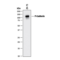
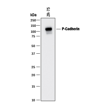
- Detection of Human P-Cadherin by Western Blot. Western blot shows lysate of ZR-75 human breast cancer cell line. PVDF membrane was probed with 1 µg/mL of Mouse Anti-Human P-Cadherin Monoclonal Antibody (Catalog # MAB861) followed by HRP-conjugated Anti-Mouse IgG Secondary Antibody (Catalog # HAF018). A specific band was detected for P-Cadherin at approximately 120 kDa (as indicated). This experiment was conducted under reducing conditions and using Immunoblot Buffer Group 1.
- Submitted by
- R&D Systems (provider)
- Main image

- Experimental details
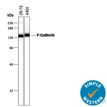
- Detection of Human P-Cadherin by Simple WesternTM. Simple Western lane view shows lysates of ZR-75 human breast cancer cell line and A431 human epithelial carcinoma cell line, loaded at 0.2 mg/mL. A specific band was detected for P-Cadherin at approximately 135 kDa (as indicated) using 10 µg/mL of Mouse Anti-Human P-Cadherin Monoclonal Antibody (Catalog # MAB861) . This experiment was conducted under reducing conditions and using the 12-230 kDa separation system.
- Submitted by
- R&D Systems (provider)
- Main image
- Experimental details
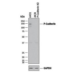
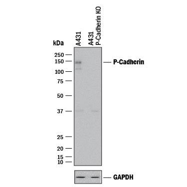
- Western Blot Shows Human P-Cadherin Specificity by Using Knockout Cell Line. Western blot shows lysates of A431 human epithelial carcinoma parental cell line and P-Cadherin knockout A431 cell line (KO). PVDF membrane was probed with 2 µg/mL of Mouse Anti-Human P-Cadherin Monoclonal Antibody (Catalog # MAB861) followed by HRP-conjugated Anti-Mouse IgG Secondary Antibody (Catalog # HAF018). A specific band was detected for P-Cadherin at approximately 150 kDa (as indicated) in the parental A431 cell line, but is not detectable in knockout A431 cell line. GAPDH (Catalog # MAB5718) is shown as a loading control. This experiment was conducted under reducing conditions and using Immunoblot Buffer Group 1.
Supportive validation
- Submitted by
- R&D Systems (provider)
- Main image
- Experimental details
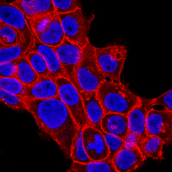
- P-Cadherin in A431 Human Cell Line. P-Cadherin was detected in immersion fixed A431 human epithelial carcinoma cell line using Mouse Anti-Human P-Cadherin Monoclonal Antibody (Catalog # MAB861) at 10 µg/mL for 3 hours at room temperature. Cells were stained using the NorthernLights™ 557-conjugated Anti-Mouse IgG Secondary Antibody (red; Catalog # NL007) and counterstained with DAPI(blue). Specific staining was localized to the cell surface. View our protocol for Fluorescent ICC Staining of Cells on Coverslips.

- Submitted by
- R&D Systems (provider)
- Main image

- Experimental details
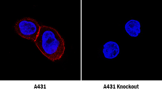
- P-Cadherin Specificity is Shown by Immunocytochemistry in Knockout Cell Line. P-Cadherin was detected in immersion fixed A431 human epithelial carcinoma cell line, wildtype (left panel) but is not detected in P-Cadherin knockout (right panel), using Mouse Anti-Human P-Cadherin Monoclonal Antibody (Catalog # MAB861) at 1 µg/mL for 3 hours at room temperature. Cells were stained using the NorthernLights 557-conjugated Anti-Mouse IgG Secondary Antibody (red; Catalog # NL007) and counterstained with DAPI (blue). Specific staining was localized to plasma membrane in wildtype cells. View our protocol for Fluorescent ICC Staining of Cells on Coverslips.
Supportive validation
- Submitted by
- R&D Systems (provider)
- Main image

- Experimental details
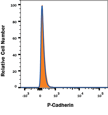
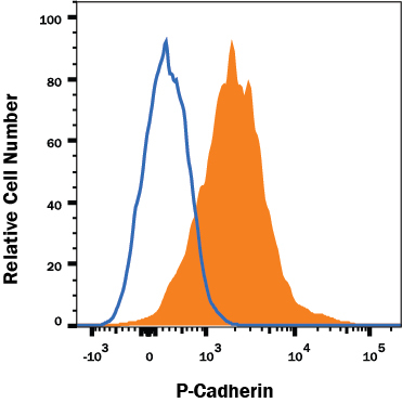
- Detection of P-Cadherin in A431 Human Cell Line by Flow Cytometry. A431 human carcinoma cell line was stained with Mouse Anti-Human P-Cadherin Monoclonal Antibody (Catalog # MAB861, filled histogram) or isotype control antibody (Catalog # MAB002, open histogram), followed by Allophycocyanin-conjugated Anti-Mouse IgG F(ab')2 Secondary Antibody (Catalog # F0101B). Cells were stained in a buffer containing Ca2+ and Mg2+. View our protocol for Staining Membrane-associated Proteins.
- Submitted by
- R&D Systems (provider)
- Main image
- Experimental details
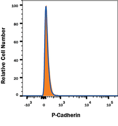
- P-Cadherin Specificity is Shown by Flow Cytometry in Knockout Cell Line. P-Cadherin knockout A431 human epithelial carcinoma cell line was stained with Mouse Anti-Human P-Cadherin Monoclonal Antibody (Catalog # MAB861, filled histogram) or isotype control antibody (Catalog # MAB002, open histogram) followed by anti-Mouse IgG PE-conjugated Secondary Antibody (Catalog # F0102B). No staining in the P-Cadherin knockout A431 cell line was observed. Cells were stained in a buffer containing Ca2+ and Mg2+. View our protocol for Staining Membrane-associated Proteins.