Explore
 Validate
 Learn
Learn Western blot
 Immunocytochemistry
Antibody data
- Antibody Data
- Antigen structure
- References [2]
- Comments [0]
- Validations
- Immunocytochemistry [1]
- Flow cytometry [1]
Submit
Validation data
Reference
Comment
Report error
- Product number
- MAB761-100 - Provider product page

- Provider
- R&D Systems
- Product name
- Human/Mouse P-Cadherin Antibody
- Antibody type
- Monoclonal
- Description
- Protein A or G purified from hybridoma culture supernatant. Detects mouse P-Cadherin in direct ELISAs and Western blots. In direct ELISAs and Western blots, no cross-reactivity with recombinant human (rh) N-Cadherin, recombinant mouse (rm) VE-Cadherin, rhCadherin-8, or rhCadherin-17 is observed.
- Reactivity
- Human, Mouse
- Host
- Rat
- Conjugate
- Unconjugated
- Antigen sequence
Q8BSL6- Isotype
- IgG
- Antibody clone number
- 106020
- Vial size
- 100 ug
- Storage
- Use a manual defrost freezer and avoid repeated freeze-thaw cycles. 12 months from date of receipt, -20 to -70 °C as supplied. 1 month, 2 to 8 °C under sterile conditions after reconstitution. 6 months, -20 to -70 °C under sterile conditions after reconstitution.
Submitted references Matrix metalloproteinase-9 deficiency attenuates diabetic nephropathy by modulation of podocyte functions and dedifferentiation.
New insights into cadherin function in epidermal sheet formation and maintenance of tissue integrity.
Li SY, Huang PH, Yang AH, Tarng DC, Yang WC, Lin CC, Chen JW, Schmid-Schönbein G, Lin SJ
Kidney international 2014 Aug;86(2):358-69
Kidney international 2014 Aug;86(2):358-69
New insights into cadherin function in epidermal sheet formation and maintenance of tissue integrity.
Tinkle CL, Pasolli HA, Stokes N, Fuchs E
Proceedings of the National Academy of Sciences of the United States of America 2008 Oct 7;105(40):15405-10
Proceedings of the National Academy of Sciences of the United States of America 2008 Oct 7;105(40):15405-10
No comments: Submit comment
Supportive validation
- Submitted by
- R&D Systems (provider)
- Main image
- Experimental details
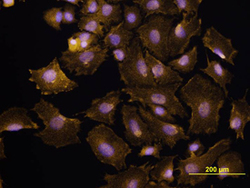
- P-Cadherin in XB2 Mouse Cell Line. P-Cadherin was detected in immersion fixed XB2 mouse teratoma keratinocyte cell line using Anti-Human/Mouse P-Cadherin Monoclonal Antibody (Catalog # MAB761) at 10 µg/mL for 3 hours at room temperature. Cells were stained using the NorthernLights™ 557-conjugated Anti-Rat IgG Secondary Antibody (yellow; Catalog # NL013) and counterstained with DAPI (blue). View our protocol for Fluorescent ICC Staining of Cells on Coverslips.

Supportive validation
- Submitted by
- R&D Systems (provider)
- Main image

- Experimental details
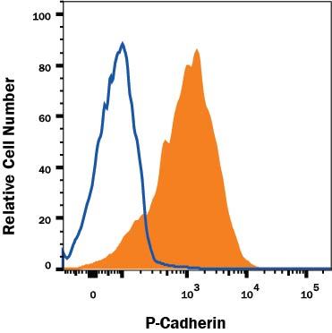
- Detection of P-Cadherin in A431 Human Cell Line by Flow Cytometry. A431 human epithelial carcinoma cell line was stained with Rat Anti-Human/Mouse P-Cadherin Monoclonal Antibody (Catalog # MAB761, filled histogram) or isotype control antibody (Catalog # MAB006, open histogram), followed by Allophycocyanin-conjugated Anti-Mouse IgG Secondary Antibody (Catalog # F0101B). Cells were stained in a buffer containing Ca2+ and Mg2+ . View our protocol for Staining Membrane-associated Proteins.