 Explore
 Validate
 Learn
Learn Western blot
Antibody data
- Antibody Data
- Antigen structure
- References [0]
- Comments [0]
- Validations
- Western blot [2]
- Immunocytochemistry [1]
- Flow cytometry [1]
Submit
Validation data
Reference
Comment
Report error
- Product number
- 14-2237-80 - Provider product page

- Provider
- Invitrogen Antibodies
- Product name
- P-Cadherin Monoclonal Antibody (CSTEM29), eBioscience™
- Antibody type
- Monoclonal
- Antigen
- Other
- Description
- This monoclonal antibody CSTEM29 reacts with human P-Cadherin, also known as Cadherin-3.
- Antibody clone number
- CSTEM29
- Concentration
- 0.5 mg/mL
No comments: Submit comment
Supportive validation
- Submitted by
- Invitrogen Antibodies (provider)
- Main image
- Experimental details
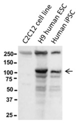
- Western blot of reduced murine C2C12 myoblast cells, H9 human ESCs, or human iPSCs using 10 µg/mL of Anti-Human P-Cadherin Purified antibody. Bands were visualized using Anti-Mouse IgG HRP.

- Submitted by
- Invitrogen Antibodies (provider)
- Main image
- Experimental details
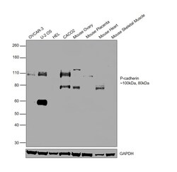
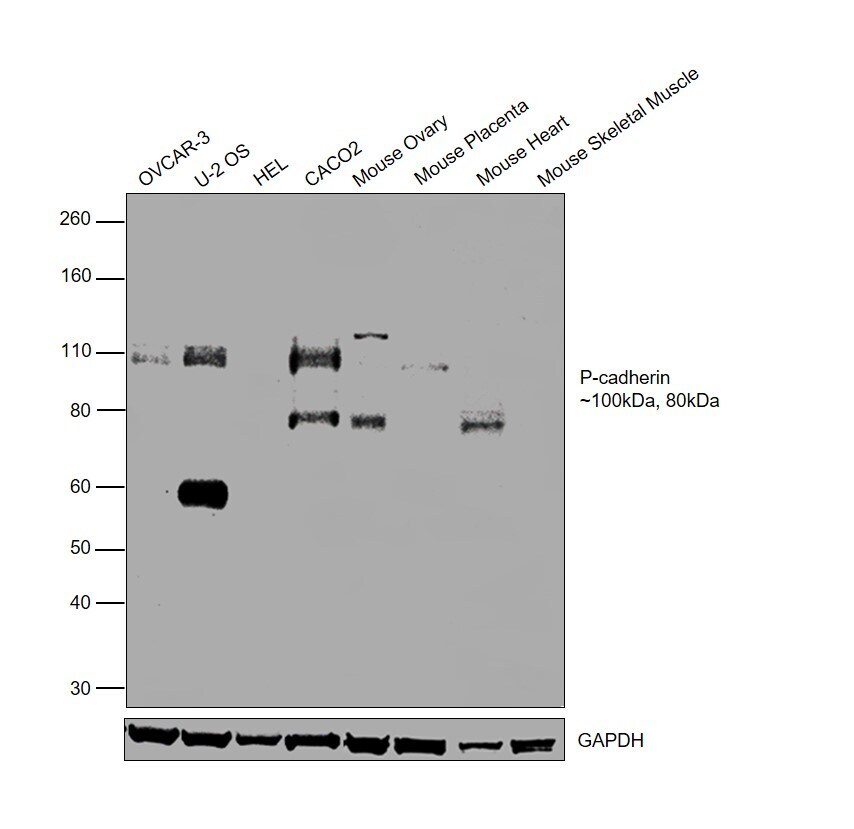
- Western blot was performed using Anti-P-Cadherin Monoclonal Antibody (CSTEM29), eBioscience™ (Product # 14-2237-82) and 100kDa and 80kDa bands corresponding to Cadherin-3 was observed across all the tested cell lines and tissues, except HEL 92.1.7 and Mouse Skeletal Muscle. Whole cell extracts (40 µg lysate) of NIH:OVCAR-3 (Lane 1), U-2 OS (Lane 2), HEL 92.1.7 (Lane 3), Caco-2 (Lane 4), Mouse Ovary (Lane 5), Mouse Placenta (Lane 6), Mouse Heart (Lane 7), Mouse Skeletal Muscle (Lane 8) were electrophoresed using NuPAGE™ 4-12% Bis-Tris Protein Gel (Product # NP0322BOX). Resolved proteins were then transferred onto a Nitrocellulose membrane (Product # IB23001) by iBlot® 2 Dry Blotting System (Product # IB21001). The blot was probed with the primary antibody (10 µg/mL) and detected by chemiluminescence with Goat anti-Mouse IgG (H+L) Superclonal™ Recombinant Secondary Antibody, HRP (Product # A28177, 1:4000 dilution) using the iBright FL 1000 (Product # A32752). Chemiluminescent detection was performed using SuperSignal™ West Dura Extended Duration Substrate (Product # 34076).
Supportive validation
- Submitted by
- Invitrogen Antibodies (provider)
- Main image
- Experimental details
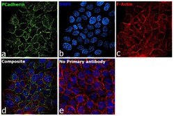
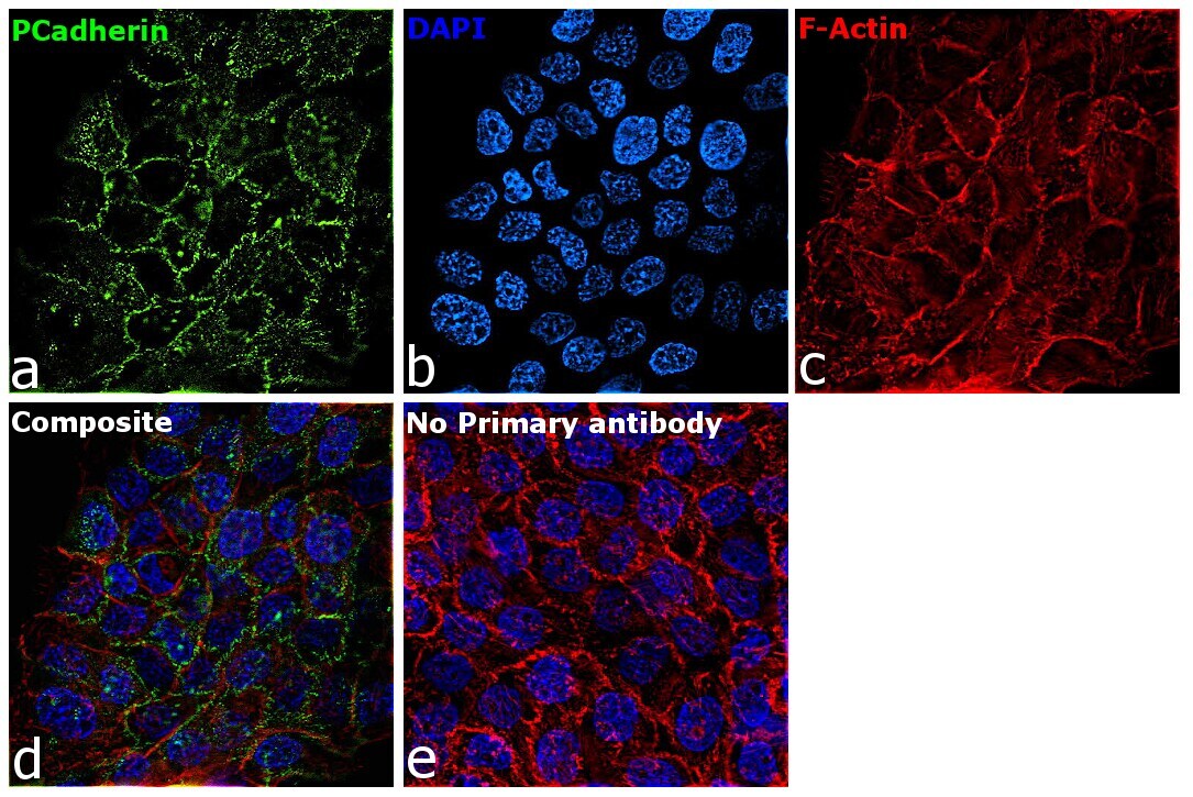
- Immunofluorescence analysis of Cadherin-3 was performed using 70% confluent log phase A-431 cells. The cells were fixed with 4% paraformaldehyde for 10 minutes, permeabilized with 0.01% Triton™ X-100 for 15 minutes, and blocked with 2% BSA for 45 minutes at room temperature. The cells were labeled with P-Cadherin Monoclonal Antibody (CSTEM29), eBioscience™ (Product # 14-2237-82) at 5µg/mL in 0.1% BSA, incubated at 4 degree celsius overnight and then labeled with Donkey anti-Mouse IgG (H+L) Highly Cross-Adsorbed Secondary Antibody, Alexa Fluor Plus 488 (Product # A32766, 1:2000 dilution), for 45 minutes at room temperature (Panel a: Green). Nuclei (Panel b: Blue) were stained with ProLong™ Diamond Antifade Mountant with DAPI (Product # P36962). F-actin (Panel c: Red) was stained with Rhodamine Phalloidin (Product # R415, 1:300 dilution). Panel d represents the merged image showing cell junction localization. Panel e represents control cells with no primary antibody to assess background. The images were captured at 60X magnification.
Supportive validation
- Submitted by
- Invitrogen Antibodies (provider)
- Main image
- Experimental details
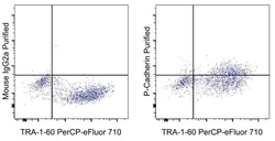
- Staining of a mixture of human iPSC and the C2C12 murine cell line with Anti-Human TRA-1-60 PerCP-eFluor 710 (Product # 46-8863-82) and 0.25 µg Mouse IgG2a K Isotype Control Purified (Product # 14-4724-82) (left) or 0.25 µg of Anti-Human P-Cadherin Purified (right) followed by F (ab')2 Anti-Mouse IgG PE (Product # 12-4010-82).
