 Explore
 Validate
 Learn
Learn Western blot
Antibody data
- Antibody Data
- Antigen structure
- References [0]
- Comments [0]
- Validations
- Western blot [2]
- Immunocytochemistry [1]
- Flow cytometry [1]
Submit
Validation data
Reference
Comment
Report error
- Product number
- 14-2237-82 - Provider product page

- Provider
- Invitrogen Antibodies
- Product name
- P-Cadherin Monoclonal Antibody (CSTEM29), eBioscience™
- Antibody type
- Monoclonal
- Antigen
- Other
- Description
- This monoclonal antibody CSTEM29 reacts with human P-Cadherin, also known as Cadherin-3. Applications Reported: This CSTEM29 antibody has been reported for use in flow cytometric analysis and immunoblot. Applications Tested: This CSTEM29 antibody has been tested by flow cytometric analysis of normal human induced pluripotent stem cells, and for western blot. For flow cytometry, this may be used at less than or equal to 0.25 µg per test. A test is defined as the amount (µg) of antibody that will stain a cell sample in a final volume of 100 µL. Cell number should be determined empirically but can range from 10^5 to 10^8 cells/test. For western blot, this can be used at less than or equal to 10 µg/mL. It is recommended that the antibody be carefully titrated for optimal performance in the assay of interest. Purity: Greater than 90%, as determined by SDS-PAGE. Aggregation: Less than 10%, as determined by HPLC. Filtration: 0.2 µm post-manufacturing filtered.
- Reactivity
- Human, Mouse
- Host
- Mouse
- Isotype
- IgG
- Antibody clone number
- CSTEM29
- Vial size
- 100 µg
- Concentration
- 0.5 mg/mL
- Storage
- 4° C, do not freeze
No comments: Submit comment
Supportive validation
- Submitted by
- Invitrogen Antibodies (provider)
- Main image
- Experimental details
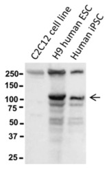
- Western blot of reduced murine C2C12 myoblast cells, H9 human ESCs, or human iPSCs using 10 µg/mL of Anti-Human P-Cadherin Purified antibody. Bands were visualized using Anti-Mouse IgG HRP.

- Submitted by
- Invitrogen Antibodies (provider)
- Main image
- Experimental details
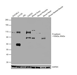
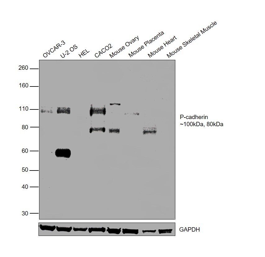
- Western blot was performed using Anti-P-Cadherin Monoclonal Antibody (CSTEM29), eBioscience™ (Product # 14-2237-82) and 100kDa and 80kDa bands corresponding to Cadherin-3 was observed across all the tested cell lines and tissues, except HEL 92.1.7 and Mouse Skeletal Muscle. Whole cell extracts (40 µg lysate) of NIH:OVCAR-3 (Lane 1), U-2 OS (Lane 2), HEL 92.1.7 (Lane 3), Caco-2 (Lane 4), Mouse Ovary (Lane 5), Mouse Placenta (Lane 6), Mouse Heart (Lane 7), Mouse Skeletal Muscle (Lane 8) were electrophoresed using NuPAGE™ 4-12% Bis-Tris Protein Gel (Product # NP0322BOX). Resolved proteins were then transferred onto a Nitrocellulose membrane (Product # IB23001) by iBlot® 2 Dry Blotting System (Product # IB21001). The blot was probed with the primary antibody (10 µg/mL) and detected by chemiluminescence with Goat anti-Mouse IgG (H+L) Superclonal™ Recombinant Secondary Antibody, HRP (Product # A28177, 1:4000 dilution) using the iBright FL 1000 (Product # A32752). Chemiluminescent detection was performed using SuperSignal™ West Dura Extended Duration Substrate (Product # 34076).
Supportive validation
- Submitted by
- Invitrogen Antibodies (provider)
- Main image
- Experimental details
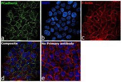
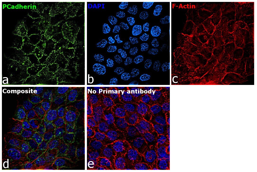
- Immunofluorescence analysis of Cadherin-3 was performed using 70% confluent log phase A-431 cells. The cells were fixed with 4% paraformaldehyde for 10 minutes, permeabilized with 0.01% Triton™ X-100 for 15 minutes, and blocked with 2% BSA for 45 minutes at room temperature. The cells were labeled with P-Cadherin Monoclonal Antibody (CSTEM29), eBioscience™ (Product # 14-2237-82) at 5µg/mL in 0.1% BSA, incubated at 4 degree celsius overnight and then labeled with Donkey anti-Mouse IgG (H+L) Highly Cross-Adsorbed Secondary Antibody, Alexa Fluor Plus 488 (Product # A32766, 1:2000 dilution), for 45 minutes at room temperature (Panel a: Green). Nuclei (Panel b: Blue) were stained with ProLong™ Diamond Antifade Mountant with DAPI (Product # P36962). F-actin (Panel c: Red) was stained with Rhodamine Phalloidin (Product # R415, 1:300 dilution). Panel d represents the merged image showing cell junction localization. Panel e represents control cells with no primary antibody to assess background. The images were captured at 60X magnification.
Supportive validation
- Submitted by
- Invitrogen Antibodies (provider)
- Main image
- Experimental details
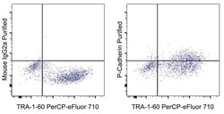
- Staining of a mixture of human iPSC and the C2C12 murine cell line with Anti-Human TRA-1-60 PerCP-eFluor 710 (Product # 46-8863-82) and 0.25 µg Mouse IgG2a K Isotype Control Purified (Product # 14-4724-82) (left) or 0.25 µg of Anti-Human P-Cadherin Purified (right) followed by F (ab')2 Anti-Mouse IgG PE (Product # 12-4010-82).
