 Explore
 Validate
 Learn
Learn Western blot
 Immunocytochemistry
Antibody data
- Antibody Data
- Antigen structure
- References [4]
- Comments [0]
- Validations
- Immunocytochemistry [1]
- Immunohistochemistry [1]
Submit
Validation data
Reference
Comment
Report error
- Product number
- 14-9873-82 - Provider product page

- Provider
- Invitrogen Antibodies
- Product name
- P-cadherin Monoclonal Antibody (12H6), eBioscience™
- Antibody type
- Monoclonal
- Antigen
- Other
- Description
- Description: The monoclonal antibody 12H6 recognizes human P-cadherin (placental-cadherin), a calcium dependent cell-cell adhesion protein. This 118 kDa transmembrane protein was originally named due to its expression in mouse placental tissue. Unfortunately, it is not expressed in human placental tissue. The function of the molecule will depend on the associations with the specific member of the cytoplasmic catenin family (alpha, beta and gamma). The specific interaction defines the strength and signaling of the cell-cell interaction. Expression is less broad than E-cadherin; P-cadherin is restricted to the basal proliferative cell layer of stratified epithelia. During development expression is found in the proliferating tissues such as hair follicle keratinocytes and the growth regions in ductal mammary tissue. There is evidence that P-cadherin can be secreted from epithelial cells during the late stages of pregnancy. P-cadherin expression is associated with poor prognosis for breast cancer while reduced expression in melanomas and squamous cell carcinomas may also result in poor prognosis. Applications Reported: This 12H6 antibody has been reported for use in western blotting, immunohistochemical staining of formalin-fixed paraffin embedded tissue sections (IHC-P), and immunocytochemical staining (ICC). Applications Tested: This 12H6 antibody has been tested immunohistochemistry on formalin-fixed paraffin embedded (FFPE) human skin with low pH antigen retrieval. This antibody can be used at less than or equal to 10 µg/mL. It is recommended that this antibody be carefully titrated for optimal performance in the assay of interest. Purity: Greater than 90%, as determined by SDS-PAGE. Aggregation: Less than 10%, as determined by HPLC. Filtration: 0.2 µm post-manufacturing filtered.
- Reactivity
- Human
- Host
- Mouse
- Isotype
- IgG
- Antibody clone number
- 12H6
- Vial size
- 100 µg
- Concentration
- 0.5 mg/mL
- Storage
- 4° C
Submitted references P-cadherin expression reduces melanoma growth, invasion, and responsiveness to growth factors in nude mice.
P-cadherin expression as a prognostic biomarker in a 3992 case tissue microarray series of breast cancer.
Functional characterization of E- and P-cadherin in invasive breast cancer cells.
P-cadherin expression in breast cancer: a review.
Jacobs K, Feys L, Vanhoecke B, Van Marck V, Bracke M
European journal of cancer prevention : the official journal of the European Cancer Prevention Organisation (ECP) 2011 May;20(3):207-16
European journal of cancer prevention : the official journal of the European Cancer Prevention Organisation (ECP) 2011 May;20(3):207-16
P-cadherin expression as a prognostic biomarker in a 3992 case tissue microarray series of breast cancer.
Turashvili G, McKinney SE, Goktepe O, Leung SC, Huntsman DG, Gelmon KA, Los G, Rejto PA, Aparicio SA
Modern pathology : an official journal of the United States and Canadian Academy of Pathology, Inc 2011 Jan;24(1):64-81
Modern pathology : an official journal of the United States and Canadian Academy of Pathology, Inc 2011 Jan;24(1):64-81
Functional characterization of E- and P-cadherin in invasive breast cancer cells.
Sarrió D, Palacios J, Hergueta-Redondo M, Gómez-López G, Cano A, Moreno-Bueno G
BMC cancer 2009 Mar 3;9:74
BMC cancer 2009 Mar 3;9:74
P-cadherin expression in breast cancer: a review.
Paredes J, Correia AL, Ribeiro AS, Albergaria A, Milanezi F, Schmitt FC
Breast cancer research : BCR 2007;9(5):214
Breast cancer research : BCR 2007;9(5):214
No comments: Submit comment
Supportive validation
- Submitted by
- Invitrogen Antibodies (provider)
- Main image

- Experimental details
- Immunofluorescence analysis of Cadherin-3 was performed using 70% confluent log phase A-431 cells. The cells were fixed with 4% paraformaldehyde for 10 minutes, permeabilized with 0.01% Triton™ X-100 for 15 minutes, and blocked with 2% BSA for 45 minutes at room temperature. The cells were labeled with P-cadherin Monoclonal Antibody (12H6), eBioscience™ (Product # 14-9873-82) at 5µg/mL in 0.1% BSA, incubated at 4 degree celsius overnight and then labeled with Donkey anti-Mouse IgG (H+L) Highly Cross-Adsorbed Secondary Antibody, Alexa Fluor Plus 488 (Product # A32766, 1:2000 dilution), for 45 minutes at room temperature (Panel a: Green). Nuclei (Panel b: Blue) were stained with ProLong™ Diamond Antifade Mountant with DAPI (Product # P36962). F-actin (Panel c: Red) was stained with Rhodamine Phalloidin (Product # R415, 1:300). Panel d represents the merged image showing cell junction localization. Panel e represents control cells with no primary antibody to assess background. The images were captured at 60X magnification.

Supportive validation
- Submitted by
- Invitrogen Antibodies (provider)
- Main image
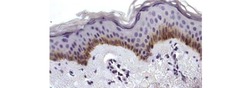
- Experimental details
- Immunohistochemistry on formalin-fixed paraffin embedded human skin using 10 µg/mL of Anti-Human P-Cadherin Purified followed by Anti-Mouse IgG Biotin, and DAB visualization.Nuclei are counterstained with hematoxylin.
