 Explore
 Validate
 Learn
Learn Flow cytometry
Antibody data
- Antibody Data
- Antigen structure
- References [0]
- Comments [0]
- Validations
- Flow cytometry [2]
Submit
Validation data
Reference
Comment
Report error
- Product number
- FAB861A - Provider product page

- Provider
- R&D Systems
- Product name
- Human P-Cadherin APC-conjugated Antibody
- Antibody type
- Monoclonal
- Description
- Protein A or G purified from hybridoma culture supernatant. Detects human P-Cadherin in direct ELISAs and Western blots. In Western blots, does not cross-react with recombinant human (rh) Cadherin-8, recombinant mouse P-Cadherin, or rhVE-Cadherin.
- Reactivity
- Human
- Host
- Mouse
- Conjugate
- Red dye
- Antigen sequence
CAA45177- Isotype
- IgG
- Antibody clone number
- 104805
- Vial size
- 100 Tests
- Storage
- Protect from light. Do not freeze. 12 months from date of receipt, 2 to 8 °C as supplied.
No comments: Submit comment
Supportive validation
- Submitted by
- R&D Systems (provider)
- Main image

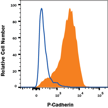
- Experimental details
- Detection of P-Cadherin in A431 Human Cell Line by Flow Cytometry. A431 human epithelial carcinoma cell line was stained with Mouse Anti-Human P-Cadherin APC-conjugated Monoclonal Antibody (Catalog # FAB861A, filled histogram) or isotype control antibody (Catalog # IC002A, open histogram). Cells were stained in a buffer containing Ca2+ and Mg2+. View our protocol for Staining Membrane-associated Proteins.
- Submitted by
- R&D Systems (provider)
- Main image
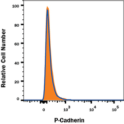
- Experimental details
- P-Cadherin Specificity is Shown by Flow Cytometry in Knockout Cell Line. P-Cadherin knockout A431 human epithelial carcinoma cell line was stained with Mouse Anti-Human APC-conjugated P-Cadherin Monoclonal Antibody (Catalog # FAB861A, filled histogram) or isotype control antibody (Catalog # IC002A, open histogram). No staining in the P-Cadherin knockout A431 cell line was observed. Cells were stained in a buffer containing Ca2+ and Mg2+. View our protocol for Staining Membrane-associated Proteins.
