 Explore
 Validate
 Learn
Learn Western blot
Antibody data
- Antibody Data
- Antigen structure
- References [7]
- Comments [0]
- Validations
- Western blot [2]
- Immunohistochemistry [1]
Submit
Validation data
Reference
Comment
Report error
- Product number
- AP6242a - Provider product page

- Provider
- Abcepta
- Proper citation
- Abgent Cat#AP6242a, RRID:AB_2301843
- Product name
- SIRT3 Antibody (C-term)
- Antibody type
- Polyclonal
- Antigen
- Synthetic peptide
- Description
- Purified Rabbit Polyclonal Antibody (Pab)
- Reactivity
- Human, Mouse
- Host
- Rabbit
- Isotype
- IgG
- Vial size
- 400 µl
- Concentration
- 0.5 mg/ml
- Storage
- Maintain refrigerated at 2-8°C for up to 6 months. For long term storage store at -20°C in small aliquots to prevent freeze-thaw cycles.
Submitted references Activation of the aryl hydrocarbon receptor sensitizes mice to nonalcoholic steatohepatitis by deactivating mitochondrial sirtuin deacetylase Sirt3.
Receptor-interacting protein (RIP) and Sirtuin-3 (SIRT3) are on opposite sides of anoikis and tumorigenesis.
FoxO1 mediates an autofeedback loop regulating SIRT1 expression.
PPARα-LXR as a novel metabolostatic signalling axis in skeletal muscle that acts to optimize substrate selection in response to nutrient status.
Sirtuin-3 (SIRT3), a novel potential therapeutic target for oral cancer.
Sirt3 blocks the cardiac hypertrophic response by augmenting Foxo3a-dependent antioxidant defense mechanisms in mice.
SIRT3 is a stress-responsive deacetylase in cardiomyocytes that protects cells from stress-mediated cell death by deacetylation of Ku70.
He J, Hu B, Shi X, Weidert ER, Lu P, Xu M, Huang M, Kelley EE, Xie W
Molecular and cellular biology 2013 May;33(10):2047-55
Molecular and cellular biology 2013 May;33(10):2047-55
Receptor-interacting protein (RIP) and Sirtuin-3 (SIRT3) are on opposite sides of anoikis and tumorigenesis.
Kamarajan P, Alhazzazi TY, Danciu T, D'silva NJ, Verdin E, Kapila YL
Cancer 2012 Dec 1;118(23):5800-10
Cancer 2012 Dec 1;118(23):5800-10
FoxO1 mediates an autofeedback loop regulating SIRT1 expression.
Xiong S, Salazar G, Patrushev N, Alexander RW
The Journal of biological chemistry 2011 Feb 18;286(7):5289-99
The Journal of biological chemistry 2011 Feb 18;286(7):5289-99
PPARα-LXR as a novel metabolostatic signalling axis in skeletal muscle that acts to optimize substrate selection in response to nutrient status.
Caton PW, Holness MJ, Bishop-Bailey D, Sugden MC
The Biochemical journal 2011 Aug 1;437(3):521-30
The Biochemical journal 2011 Aug 1;437(3):521-30
Sirtuin-3 (SIRT3), a novel potential therapeutic target for oral cancer.
Alhazzazi TY, Kamarajan P, Joo N, Huang JY, Verdin E, D'Silva NJ, Kapila YL
Cancer 2011 Apr 15;117(8):1670-8
Cancer 2011 Apr 15;117(8):1670-8
Sirt3 blocks the cardiac hypertrophic response by augmenting Foxo3a-dependent antioxidant defense mechanisms in mice.
Sundaresan NR, Gupta M, Kim G, Rajamohan SB, Isbatan A, Gupta MP
The Journal of clinical investigation 2009 Sep;119(9):2758-71
The Journal of clinical investigation 2009 Sep;119(9):2758-71
SIRT3 is a stress-responsive deacetylase in cardiomyocytes that protects cells from stress-mediated cell death by deacetylation of Ku70.
Sundaresan NR, Samant SA, Pillai VB, Rajamohan SB, Gupta MP
Molecular and cellular biology 2008 Oct;28(20):6384-401
Molecular and cellular biology 2008 Oct;28(20):6384-401
No comments: Submit comment
Supportive validation
- Submitted by
- Abcepta (provider)
- Main image
- Experimental details
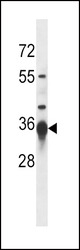
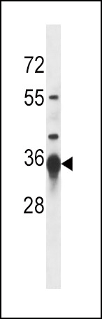
- Sirt3 Antibody (G265) (Cat. #AP6242a) western blot analysis in mouse kidney tissue lysates (35ug/lane).This demonstrates the Sirt3 antibody detected the Sirt3 protein (arrow).
- Primary Ab dilution
- 1:1000
- Submitted by
- Abcepta (provider)
- Main image
- Experimental details
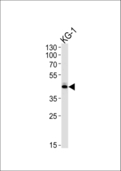
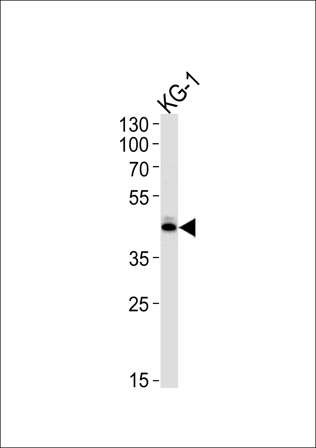
- SIRT3 Antibody (C-term) (Cat.# AP6242a) western blot analysis in KG-1 cell line lysates (35ug/lane).This demonstrates the SIRT3 antibody detected the SIRT3 protein (arrow).
- Primary Ab dilution
- 1:1000
Supportive validation
- Submitted by
- Abcepta (provider)
- Main image
- Experimental details
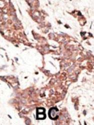
- "Formalin-fixed and paraffin-embedded human cancer tissue reacted with the primary antibody, which was peroxidase-conjugated to the secondary antibody, followed by AEC staining. This data demonstrates the use of this antibody for immunohistochemistry; clinical relevance has not been evaluated. BC = breast carcinoma; HC = hepatocarcinoma."
- Primary Ab dilution
- 1:50~100
