Explore
 Validate
 Learn
Learn Western blot
 Immunocytochemistry
Antibody data
- Antibody Data
- Antigen structure
- References [0]
- Comments [0]
- Validations
- Western blot [1]
- Other assay [1]
Submit
Validation data
Reference
Comment
Report error
- Product number
- PA5-19732 - Provider product page

- Provider
- Invitrogen Antibodies
- Product name
- Anti-SIRT3 Polyclonal Antibody
- Antibody type
- Polyclonal
- Antigen
- Synthetic peptide
- Description
- PA5-19732 targets SIRT3 in IF and WB applications and shows reactivity with Human samples. The PA5-19732 immunogen is synthetic peptide conjugated to KLH derived from within residues 50 - 150 of Human SIRT3. PA5-19732 detects SIRT3 which has a predicted molecular weight of approximately 50 kDa. Store antibody at 4ºC for 1-2 weeks. For long-term storage, store at -20ºC.
- Reactivity
- Human
- Host
- Rabbit
- Isotype
- IgG
- Vial size
- 100 µg
- Concentration
- 0.2 mg/mL
- Storage
- Store at 4°C short term. For long term storage, store at -20°C, avoiding freeze/thaw cycles.
No comments: Submit comment
Supportive validation
- Submitted by
- Invitrogen Antibodies (provider)
- Main image
- Experimental details
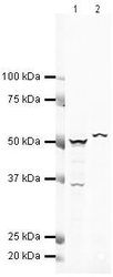
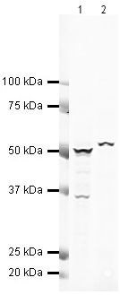
- Western blot analysis of Human Liver Tissue Lysate using Product # PA5-19732, SIRT3 primary antibody at a dilution of 1 µg/mL (lane 1). Staining of Human Lymph Node Tissue Lysate at a dilution of 1 µg/mL (lane 2). Blot treated with a secondary IR Dye680-conjugated Goat polyclonal anti-Rabbit antibody was used at a dilution of 1:10000.
Supportive validation
- Submitted by
- Invitrogen Antibodies (provider)
- Main image
- Experimental details
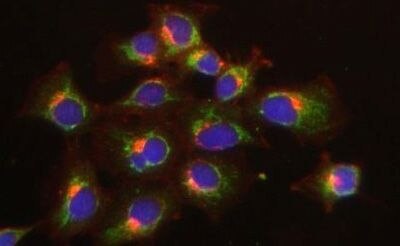
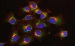
- Immunofluorescent staining of HeLa cells using Product # PA5-19732, anti-SIRT3 antibody. The cells were fixed with methanol (100%) for 5 minutes, permabilised with TBS-T (20mins), BSA (1%), normal goat serum (10%) and glycine (0.3 M) in 0.1% PBS-Tween for 1 hour and exposed to the primary antibody at a concentration of 1 µg/mL for 1 hour at room temp. The secondary antibody was a 448 fluorescence conjugated Goat anti-rabbit IgG (green) at a dilution of 1:1000. A WGA- 594 fluorescent conjugated stain was used to label plasma membranes (red) and the nuclei stain was DAPI (blue).