Explore
 Validate
 Learn
Learn Western blot
 Immunocytochemistry
Antibody data
- Antibody Data
- Antigen structure
- References [9]
- Comments [0]
- Validations
- Immunocytochemistry [1]
- Immunohistochemistry [1]
Submit
Validation data
Reference
Comment
Report error
- Product number
- HPA006028 - Provider product page

- Provider
- Atlas Antibodies
- Proper citation
- Atlas Antibodies Cat#HPA006028, RRID:AB_1078858
- Product name
- Anti-FHL2
- Antibody type
- Polyclonal
- Description
- Polyclonal Antibody against Human FHL2, Gene description: four and a half LIM domains 2, Alternative Gene Names: DRAL, SLIM3, Validated applications: WB, IHC, ICC, Uniprot ID: Q14192, Storage: Store at +4°C for short term storage. Long time storage is recommended at -20°C.
- Reactivity
- Human, Mouse, Rat
- Host
- Rabbit
- Conjugate
- Unconjugated
- Isotype
- IgG
- Vial size
- 100 µl
- Concentration
- 0.1 mg/ml
- Storage
- Store at +4°C for short term storage. Long time storage is recommended at -20°C.
- Handling
- The antibody solution should be gently mixed before use.
Submitted references FHL2 expression by cancer-associated fibroblasts promotes metastasis and angiogenesis in lung adenocarcinoma.
Dormancy-inducing 3D engineered matrix uncovers mechanosensitive and drug-protective FHL2-p21 signaling axis
FHL2 anchors mitochondria to actin and adapts mitochondrial dynamics to glucose supply
A Novel Model for Nephrotic Syndrome Reveals Associated Dysbiosis of the Gut Microbiome and Extramedullary Hematopoiesis.
Matrix mechanics controls FHL2 movement to the nucleus to activate p21 expression
Immunofluorescence and fluorescent-protein tagging show high correlation for protein localization in mammalian cells
Systematic validation of antibody binding and protein subcellular localization using siRNA and confocal microscopy
A global view of protein expression in human cells, tissues, and organs
Kanzaki R, Reid S, Bolivar P, Sjölund J, Staaf J, Larsson S, Shintani Y, Pietras K
International journal of cancer 2025 Jan 15;156(2):431-446
International journal of cancer 2025 Jan 15;156(2):431-446
Dormancy-inducing 3D engineered matrix uncovers mechanosensitive and drug-protective FHL2-p21 signaling axis
Bakhshandeh S, Heras U, Taïeb H, Varadarajan A, Lissek S, Hücker S, Lu X, Garske D, Young S, Abaurrea A, Caffarel M, Riestra A, Bragado P, Contzen J, Gossen M, Kirsch S, Warfsmann J, Honarnejad K, Klein C, Cipitria A
Science Advances 2024;10(45)
Science Advances 2024;10(45)
Seetharaman S, Devany J, Kim H, van Bodegraven E, Chmiel T, Tzu-Pin S, Chou W, Fang Y, Gardel M
2024
2024
FHL2 anchors mitochondria to actin and adapts mitochondrial dynamics to glucose supply
Basu H, Pekkurnaz G, Falk J, Wei W, Chin M, Steen J, Schwarz T
Journal of Cell Biology 2021;220(10)
Journal of Cell Biology 2021;220(10)
A Novel Model for Nephrotic Syndrome Reveals Associated Dysbiosis of the Gut Microbiome and Extramedullary Hematopoiesis.
Maier JI, Rogg M, Helmstädter M, Sammarco A, Walz G, Werner M, Schell C
Cells 2021 Jun 15;10(6)
Cells 2021 Jun 15;10(6)
Matrix mechanics controls FHL2 movement to the nucleus to activate p21 expression
Nakazawa N, Sathe A, Shivashankar G, Sheetz M
Proceedings of the National Academy of Sciences 2016;113(44)
Proceedings of the National Academy of Sciences 2016;113(44)
Immunofluorescence and fluorescent-protein tagging show high correlation for protein localization in mammalian cells
Stadler C, Rexhepaj E, Singan V, Murphy R, Pepperkok R, Uhlén M, Simpson J, Lundberg E
Nature Methods 2013;10(4):315-323
Nature Methods 2013;10(4):315-323
Systematic validation of antibody binding and protein subcellular localization using siRNA and confocal microscopy
Stadler C, Hjelmare M, Neumann B, Jonasson K, Pepperkok R, Uhlén M, Lundberg E
Journal of Proteomics 2012;75(7):2236-2251
Journal of Proteomics 2012;75(7):2236-2251
A global view of protein expression in human cells, tissues, and organs
Pontén F, Gry M, Fagerberg L, Lundberg E, Asplund A, Berglund L, Oksvold P, Björling E, Hober S, Kampf C, Navani S, Nilsson P, Ottosson J, Persson A, Wernérus H, Wester K, Uhlén M
Molecular Systems Biology 2009;5(1)
Molecular Systems Biology 2009;5(1)
No comments: Submit comment
Supportive validation
- Submitted by
- Atlas Antibodies (provider)
- Main image
- Experimental details
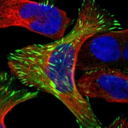
- Immunofluorescent staining of human cell line U-2 OS shows localization to actin filaments & focal adhesion sites.
- Sample type
- Human

Supportive validation
- Submitted by
- Atlas Antibodies (provider)
- Enhanced method
- Orthogonal validation
- Main image

- Experimental details
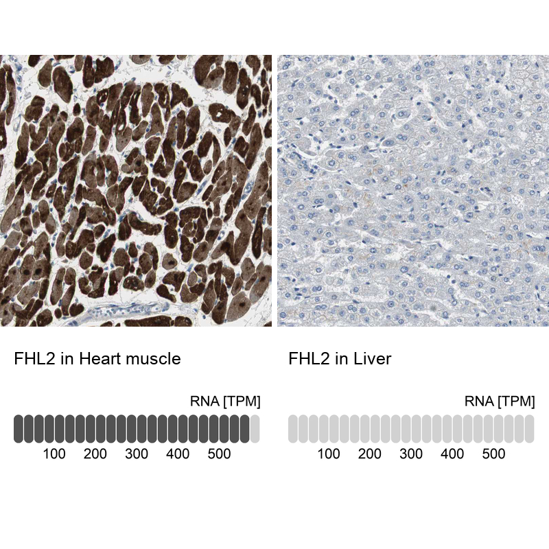
- Immunohistochemistry analysis in human heart muscle and liver tissues using HPA006028 antibody. Corresponding FHL2 RNA-seq data are presented for the same tissues.
- Sample type
- Human
- Protocol
- Protocol