Explore
 Validate
 Learn
Learn Western blot
 Immunohistochemistry
Antibody data
- Antibody Data
- Antigen structure
- References [6]
- Comments [0]
- Validations
- Western blot [1]
Submit
Validation data
Reference
Comment
Report error
- Product number
- HPA002132 - Provider product page

- Provider
- Atlas Antibodies
- Proper citation
- Atlas Antibodies Cat#HPA002132, RRID:AB_1078807
- Product name
- Anti-ESYT2
- Antibody type
- Polyclonal
- Description
- Polyclonal Antibody against Human ESYT2, Gene description: extended synaptotagmin-like protein 2, Alternative Gene Names: CHR2SYT, FAM62B, KIAA1228, Validated applications: WB, IHC, Uniprot ID: A0FGR8, Storage: Store at +4°C for short term storage. Long time storage is recommended at -20°C.
- Reactivity
- Human
- Host
- Rabbit
- Conjugate
- Unconjugated
- Isotype
- IgG
- Vial size
- 100 µl
- Concentration
- 0.2 mg/ml
- Storage
- Store at +4°C for short term storage. Long time storage is recommended at -20°C.
- Handling
- The antibody solution should be gently mixed before use.
Submitted references Endoplasmic reticulum–plasma membrane contact gradients direct cell migration
Extended-Synaptotagmin-1 and -2 control T cell signaling and function.
Cohesin-independent STAG proteins interact with RNA and R-loops and promote complex loading
Chemoproteomic profiling reveals cellular targets of nitro-fatty acids
Triggered Ca 2+ influx is required for extended synaptotagmin 1‐induced ER ‐plasma membrane tethering
Three-dimensional architecture of extended synaptotagmin-mediated endoplasmic reticulum–plasma membrane contact sites
Gong B, Johnston J, Thiemicke A, de Marco A, Meyer T
Nature 2024;631(8020):415-423
Nature 2024;631(8020):415-423
Extended-Synaptotagmin-1 and -2 control T cell signaling and function.
Benavides N, Giraudo CG
EMBO reports 2024 Jan;25(1):286-303
EMBO reports 2024 Jan;25(1):286-303
Cohesin-independent STAG proteins interact with RNA and R-loops and promote complex loading
Li Y, Porter H, Neguembor M, Beltran M, Varsally W, Martin L, Cornejo M, Pezić D, Bhamra A, Surinova S, Jenner R, Cosma M, Hadjur S
eLife 2023;12
eLife 2023;12
Chemoproteomic profiling reveals cellular targets of nitro-fatty acids
Fang M, Huang K, Tu W, Chen Y, Pan P, Hsiao W, Ke Y, Tsou L, Zhang M
Redox Biology 2021;46
Redox Biology 2021;46
Triggered Ca 2+ influx is required for extended synaptotagmin 1‐induced ER ‐plasma membrane tethering
Idevall‐Hagren O, Lü A, Xie B, De Camilli P
The EMBO Journal 2015;34(17):2291-2305
The EMBO Journal 2015;34(17):2291-2305
Three-dimensional architecture of extended synaptotagmin-mediated endoplasmic reticulum–plasma membrane contact sites
Fernández-Busnadiego R, Saheki Y, De Camilli P
Proceedings of the National Academy of Sciences 2015;112(16)
Proceedings of the National Academy of Sciences 2015;112(16)
No comments: Submit comment
Enhanced validation
- Submitted by
- Atlas Antibodies (provider)
- Enhanced method
- Genetic validation
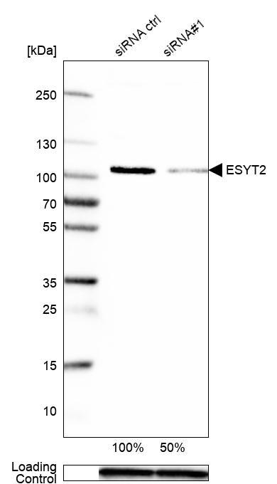
- Main image

- Experimental details
- Western blot analysis in MCF-7 cells transfected with control siRNA, target specific siRNA probe #1, using Anti-ESYT2 antibody. Remaining relative intensity is presented. Loading control: Anti-GAPDH.
- Sample type
- Human
- Protocol
- Protocol