Explore
 Validate
 Learn
Learn Western blot
Antibody data
- Antibody Data
- Antigen structure
- References [2]
- Comments [0]
- Validations
- Western blot [6]
- Immunocytochemistry [4]
- Immunoprecipitation [1]
- Immunohistochemistry [2]
- Other assay [2]
Submit
Validation data
Reference
Comment
Report error
- Product number
- PA1-112 - Provider product page

- Provider
- Invitrogen Antibodies
- Product name
- PAX8 Polyclonal Antibody
- Antibody type
- Polyclonal
- Antigen
- Purifed from natural sources
- Description
- Western blot analysis of PA1-112 detects a prominent ~48 kDa protein in human adenocarcinoma SKOV-3 and SV40 transformed African green monkey (Cercopithecus aethiops) kidney cells. In SKOV-3 cells, 2 additional unknown bands at (~20 kD) and at ~150 kDa are also detected. Specificity of the antibody was confirmed in HeLa and 293T cells overexpressing full lenght PAX8. The 48 kDa band is enriched in immunoprecipitations from Cos7 lysate, however additional unknown high molecular weight bands are also enriched and detected by PA1-112.
- Reactivity
- Human
- Host
- Rabbit
- Isotype
- IgG
- Vial size
- 100 μg
- Concentration
- 1 mg/mL
- Storage
- -20°C
Submitted references PAX8 expression in high-grade serous ovarian cancer positively regulates attachment to ECM via Integrin β3.
Candidate genes and pathways downstream of PAX8 involved in ovarian high-grade serous carcinoma.
Soriano AA, de Cristofaro T, Di Palma T, Dotolo S, Gokulnath P, Izzo A, Calì G, Facchiano A, Zannini M
Cancer cell international 2019;19:303
Cancer cell international 2019;19:303
Candidate genes and pathways downstream of PAX8 involved in ovarian high-grade serous carcinoma.
de Cristofaro T, Di Palma T, Soriano AA, Monticelli A, Affinito O, Cocozza S, Zannini M
Oncotarget 2016 Jul 5;7(27):41929-41947
Oncotarget 2016 Jul 5;7(27):41929-41947
No comments: Submit comment
Supportive validation
- Submitted by
- Invitrogen Antibodies (provider)
- Main image

- Experimental details
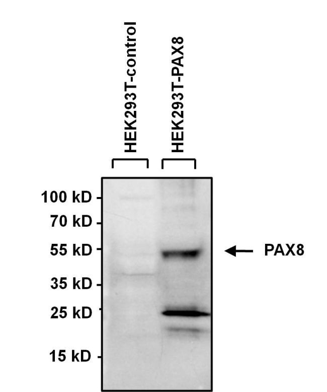
- Western blot analysis of PAX8 was performed by loading 40 µg of HEK293 lysate overexpressing PAX8 (right lane) or empty vector control (left lane) and 10 µL PageRuler Prestained Protein Ladder (Product # 26616) onto a 4-20% Tris-HCl polyacrylamide gel. Proteins were transferred to a PVDF membrane and blocked with 5% BSA/TBST for at least 1 hour. The membrane was probed with a PAX8 polyclonal antibody (Product # PA1-112) at a dilution of 1:1000 overnight at 4°C on a rocking platform, washed in TBS-0.1%Tween-20, and probed with a goat anti-rabbit HRP secondary antibody (Product # 32460) at a dilution of 1:500 (20 ng/mL) for at least one hour. Chemiluminescent detection was performed using SuperSignal West Pico (Product # 34080).
- Submitted by
- Invitrogen Antibodies (provider)
- Main image
- Experimental details
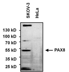
- Western blot analysis of PAX8 was performed by loading 75 µg of various whole cell lysates and 10 µL of PageRuler Prestained Protein Ladder (Product # 26616) onto a 4-20% Tris-HCl polyacrylamide gel. Proteins were transferred to a PVDF membrane and blocked with 5% BSA/TBST for at least 1 hour. The membrane was probed with a PAX8 polyclonal antibody (Product # PA1-112) at a dilution of 1:1000 overnight at 4°C on a rocking platform, washed in TBS-0.1%Tween-20, and probed with a goat anti-rabbit HRP secondary antibody (Product # 32460) at a dilution of 1:500 (20 ng/mL) for at least one hour. Chemiluminescent detection was performed using SuperSignal West Pico (Product # 34080).

- Submitted by
- Invitrogen Antibodies (provider)
- Main image
- Experimental details
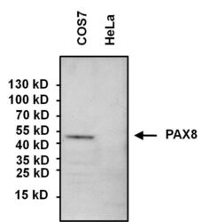
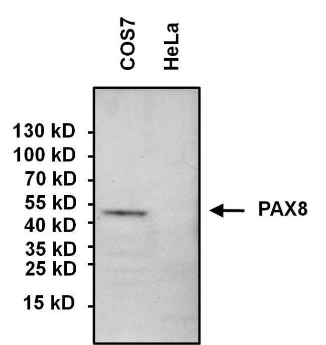
- Western blot analysis of PAX8 was performed by loading 60 µg of COS7 whole cell lysate (left lane), negative control HeLa lysate (right lane), and 10 µL PageRuler Prestained Protein Ladder (Product # 26616) onto a 4-20% Tris-HCl polyacrylamide gel. Proteins were transferred to a PVDF membrane and blocked with 5% BSA/TBST for at least 1 hour. The membrane was probed with a PAX8 polyclonal antibody (Product # PA1-112) at a dilution of 1:1000 overnight at 4°C on a rocking platform, washed in TBS-0.1%Tween-20, and probed with a goat anti-rabbit HRP secondary antibody (Product # 32460) at a dilution of 1:500 (20 ng/mL) for at least one hour. Chemiluminescent detection was performed using SuperSignal West Pico (Product # 34080).
- Submitted by
- Invitrogen Antibodies (provider)
- Main image
- Experimental details
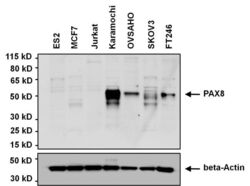
- Western blot analysis of PAX8 was performed by loading 20 µg of the indicated whole cell lysates onto a 4-12% Novex Bis-Tris polyacrylamide gel. Proteins were transferred to a nitrocellulose membrane and blocked with 5% milk in PBST for 1 hour. The membrane was probed with a PAX8 polyclonal antibody (Product # PA1-112) at a dilution of 1:500 overnight at 4C on a rocking platform, washed in PBST, and probed with a goat anti-rabbit IgG secondary antibody. The samples were also probed with a beta-actin antibody to control for equal protein loading. Chemiluminescent detection was performed using ECL Plus substrate (Product # 32132). The lysates were: ES2 ovarian clear cell carcinoma, MCF7 breast cancer, Jurkat T cell leukemia, Kuramochi high grade serious ovarian cancer, OVSAHO high grade serious ovarian carcinoma, SKOV3 ovarian adenocarcinoma, and FT246 immortalized fallopian tube epithelium cells. Data courtesy of the Innovators Program.

- Submitted by
- Invitrogen Antibodies (provider)
- Main image

- Experimental details
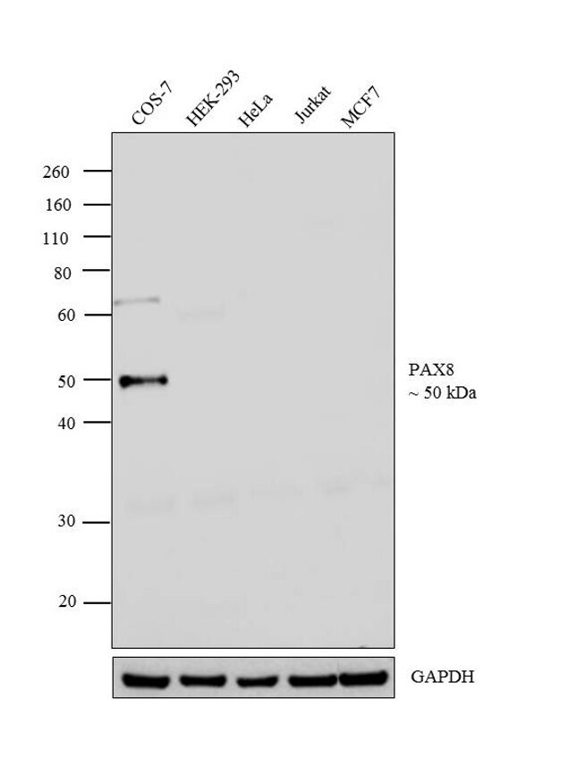
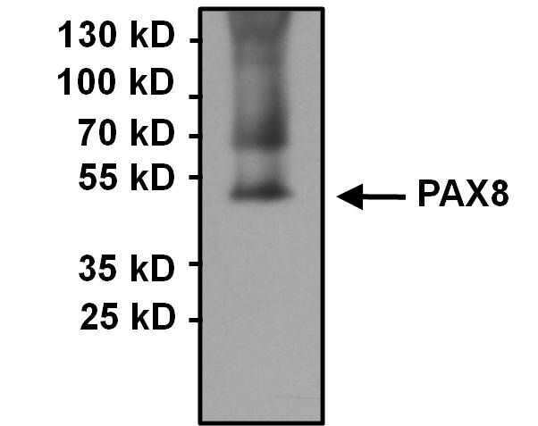
- Western blot analysis was performed on modified whole cell extracts (1% SDS) (30 µg lysate) of COS-7 (Lane 1), HEK-293 (Lane 2), HeLa (Lane 3), Jurkat (Lane 4) and MCF7 (Lane 5). The blot was probed with Anti-PAX8 Polyclonal Antibody (Product # PA1-112, 1:500 dilution) and detected by chemiluminescence using Goat anti-Rabbit IgG (Heavy Chain) Superclonal™ Secondary Antibody, HRP conjugate (Product # A27036, 0.25 µg/mL, 1:4000 dilution). A 50 kDa band corresponding to PAX8 was detected in COS-7 and not in HEK-293, HeLa, Jurkat and MCF7 which are reported negative for PAX8.
- Submitted by
- Invitrogen Antibodies (provider)
- Main image

- Experimental details
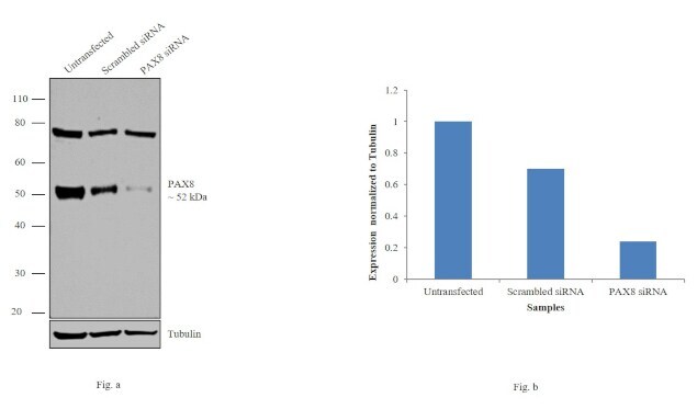
- Knockdown of PAX8 was achieved by transfecting COS-7 cells with PAX8 specific siRNAs (Silencer® select Product # n304366, s15404). Western blot analysis (Fig. a) was performed using whole cell extracts from the PAX8 knockdown cells (lane 3), non-specific scrambled siRNA transfected cells (lane 2) and untransfected cells (lane 1). The blots were probed with Anti-PAX8 Polyclonal Antibody (Product # PA1-112, 1:1000 dilution) and Goat anti-Rabbit IgG (Heavy Chain) Superclonal™ Secondary Antibody, HRP conjugate (Product # A27036, 0.25 µg/mL, 1:4000 dilution). Densitometric analysis of this western blot is shown in histogram (Fig. b). Decrease in signal upon siRNA mediated knock down confirms that antibody is specific to PAX8.
Supportive validation
- Submitted by
- Invitrogen Antibodies (provider)
- Main image

- Experimental details
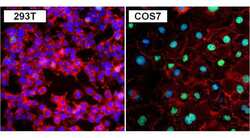
- Immunofluorescent analysis of PAX8 (green) in 293T (left panel) and COS7 cells (right panel). Formalin fixed cells were permeabilized with 0.1% Triton X-100 in TBS for 10 minutes at room temperature. Cells were blocked with 1% Blocker BSA (Product # 37525) for 15 minutes at room temperature. Cells were probed with a PAX8 polyclonal antibody (Product # PA1-112) at a dilution of 1:400 for at least 1 hour at room temperature, washed with PBS, and incubated with a DyLight 488-conjugated goat anti-rabbit IgG secondary antibody (Product # 35552) at a dilution of 1:400 for 30 minutes at room temperature. F-Actin (red) was stained with DyLight-554 Phalloidin (Product # 21834) and nuclei (blue) were stained with Hoechst 33342 dye (Product # 62249). Images were taken on a Thermo Scientific ToxInsight Instrument at 20X magnification.

- Submitted by
- Invitrogen Antibodies (provider)
- Main image
- Experimental details
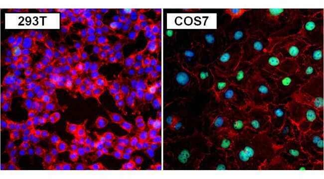
- Immunofluorescent analysis of PAX8 (green) in 293T (left panel) and COS7 cells (right panel). Formalin fixed cells were permeabilized with 0.1% Triton X-100 in TBS for 10 minutes at room temperature. Cells were blocked with 1% Blocker BSA (Product # 37525) for 15 minutes at room temperature. Cells were probed with a PAX8 polyclonal antibody (Product # PA1-112) at a dilution of 1:400 for at least 1 hour at room temperature, washed with PBS, and incubated with a DyLight 488-conjugated goat anti-rabbit IgG secondary antibody (Product # 35552) at a dilution of 1:400 for 30 minutes at room temperature. F-Actin (red) was stained with DyLight-554 Phalloidin (Product # 21834) and nuclei (blue) were stained with Hoechst 33342 dye (Product # 62249). Images were taken on a Thermo Scientific ToxInsight Instrument at 20X magnification.
- Submitted by
- Invitrogen Antibodies (provider)
- Main image

- Experimental details
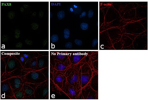
- Immunofluorescence analysis of PAX8 was performed using 70% confluent log phase Cos-7 cells. The cells were fixed with 4% paraformaldehyde for 10 minutes, permeabilized with 0.1% Triton™ X-100 for 15 minutes, and blocked with 1% BSA for 1 hour at room temperature. The cells were labeled with PAX8 Polyclonal Antibody (Product # PA1-112) at 1:100 dilution in 0.1% BSA, incubated at 4 degree Celsius overnight and then labeled with Goat anti-Rabbit IgG (H+L) Superclonal™ Secondary Antibody, Alexa Fluor® 488 conjugate (Product # A27034) at a dilution of 1:2000 for 45 minutes at room temperature (Panel a: green). Nuclei (Panel b: blue) were stained with ProLong™ Diamond Antifade Mountant with DAPI (Product # P36962). F-actin (Panel c: red) was stained with Rhodamine Phalloidin (Product # R415, 1:300). Panel d represents the merged image showing Nuclear localization. Panel e represents control cells with no primary antibody to assess background. The images were captured at 60X magnification.
- Submitted by
- Invitrogen Antibodies (provider)
- Main image

- Experimental details
- Immunofluorescence analysis of PAX8 was performed using 70% confluent log phase Cos-7 cells. The cells were fixed with 4% paraformaldehyde for 10 minutes, permeabilized with 0.1% Triton™ X-100 for 15 minutes, and blocked with 1% BSA for 1 hour at room temperature. The cells were labeled with PAX8 Polyclonal Antibody (Product # PA1-112) at 1:100 dilution in 0.1% BSA, incubated at 4 degree Celsius overnight and then labeled with Goat anti-Rabbit IgG (Heavy Chain) Superclonal™ Secondary Antibody, Alexa Fluor® 488 conjugate (Product # A27034) at a dilution of 1:2000 for 45 minutes at room temperature (Panel a: green). Nuclei (Panel b: blue) were stained with ProLong™ Diamond Antifade Mountant with DAPI (Product # P36962). F-actin (Panel c: red) was stained with Rhodamine Phalloidin (Product # R415, 1:300). Panel d represents the merged image showing Nuclear localization. Panel e represents control cells with no primary antibody to assess background. The images were captured at 60X magnification.

Supportive validation
- Submitted by
- Invitrogen Antibodies (provider)
- Main image

- Experimental details
- Immunoprecipitation of PAX8 was performed using COS7 whole cell lysate. Antigen-antibody complexes were formed by incubating 500 µg of lysate with 3 µg of a PAX8 polyclonal antibody (Product # PA1-112) overnight on a rocking platform at 4°C. The immune complexes were captured on 50 µL Protein A/G Agarose (Product # 20421), washed extensively, and eluted with 5X Lane Marker Reducing Sample Buffer (Product # 39000). Samples was resolved on a 4-20% Tris-HCl polyacrylamide gel, transferred to a PVDF membrane, and blocked with 5% BSA/TBS-0.1%Tween-20 for 1 hour. The membrane was probed with a PAX8 polyclonal antibody (Product # PA1-112) at a dilution of 1:1000 overnight rotating at 4°C. Membranes were washed in TBST, and probed with Clean-Blot IP Detection Reagent (Product # 21230) at a dilution of 1:1000 for at least one hour. Chemiluminescent detection was performed using SuperSignal West Pico (Product # 34080).

Supportive validation
- Submitted by
- Invitrogen Antibodies (provider)
- Main image
- Experimental details
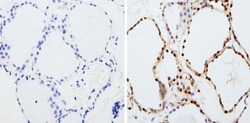
- Immunohistochemistry analysis of PAX8 showing staining in the nucleus of paraffin-embedded human thyroid tissue (right) compared to a negative control without primary antibody (left). To expose target proteins, antigen retrieval was performed using 10mM sodium citrate (pH 6.0), microwaved for 8-15 min. Following antigen retrieval, tissues were blocked in 3% H2O2-methanol for 15 min at room temperature, washed with ddH2O and PBS, and then probed with a PAX8 polyclonal antibody (Product # PA1-112) diluted in 3% BSA-PBS at a dilution of 1:200 overnight at 4°C in a humidified chamber. Tissues were washed extensively in PBST and detection was performed using an HRP-conjugated secondary antibody followed by colorimetric detection using a DAB kit. Tissues were counterstained with hematoxylin and dehydrated with ethanol and xylene to prep for mounting.
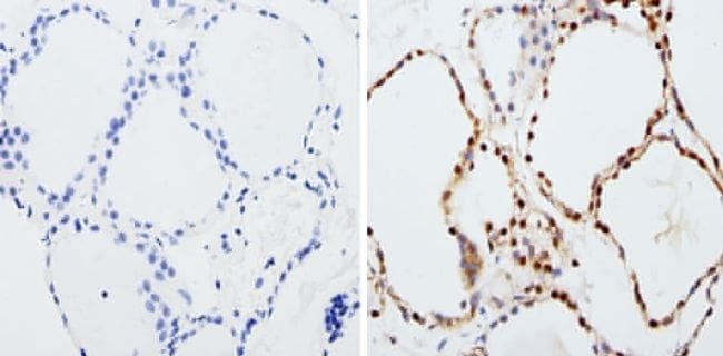
- Submitted by
- Invitrogen Antibodies (provider)
- Main image

- Experimental details
- Immunohistochemistry analysis of PAX8 showing staining in the nucleus of paraffin-embedded human thyroid tissue (right) compared to a negative control without primary antibody (left). To expose target proteins, antigen retrieval was performed using 10mM sodium citrate (pH 6.0), microwaved for 8-15 min. Following antigen retrieval, tissues were blocked in 3% H2O2-methanol for 15 min at room temperature, washed with ddH2O and PBS, and then probed with a PAX8 polyclonal antibody (Product # PA1-112) diluted in 3% BSA-PBS at a dilution of 1:200 overnight at 4°C in a humidified chamber. Tissues were washed extensively in PBST and detection was performed using an HRP-conjugated secondary antibody followed by colorimetric detection using a DAB kit. Tissues were counterstained with hematoxylin and dehydrated with ethanol and xylene to prep for mounting.

Supportive validation
- Submitted by
- Invitrogen Antibodies (provider)
- Main image

- Experimental details
- Immunoprecipitation of PAX8 was performed using COS7 whole cell lysate. Antigen-antibody complexes were formed by incubating 500 µg of lysate with 3 µg of a PAX8 polyclonal antibody (Product # PA1-112) overnight on a rocking platform at 4øC. The immune complexes were captured on 50 µL Protein A/G Agarose (Product # 20421), washed extensively, and eluted with 5X Lane Marker Reducing Sample Buffer (Product # 39000). Samples was resolved on a 4-20% Tris-HCl polyacrylamide gel, transferred to a PVDF membrane, and blocked with 5% BSA/TBS-0.1%Tween-20 for 1 hour. The membrane was probed with a PAX8 polyclonal antibody (Product # PA1-112) at a dilution of 1:1000 overnight rotating at 4øC. Membranes were washed in TBST, and probed with Clean-Blot IP Detection Reagent (Product # 21230) at a dilution of 1:1000 for at least one hour. Chemiluminescent detection was performed using SuperSignal West Pico (Product # 34080).
- Submitted by
- Invitrogen Antibodies (provider)
- Main image

- Experimental details
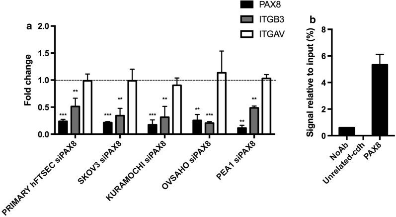
- Fig. 3 PAX8 regulates the expression of the ITGB3 gene. a PAX8, ITGB3 and ITGAV expression levels were measured by qRT-PCR in Primary hFTSEC, SKOV3, KURAMOCHI, OVSAHO and PEA1 48 h after RNAi of PAX8. The values are mean +- SD of three independent experiments in duplicate, normalized by the expression of ABL and expressed as fold change with respect to the siCTR cells, whose value was set at 1.0. p -value was calculated by t -test 0.001