Explore
 Validate
 Learn
Learn Western blot
Antibody data
- Antibody Data
- Antigen structure
- References [0]
- Comments [0]
- Validations
- Western blot [3]
- Immunocytochemistry [1]
- Immunoprecipitation [1]
- Immunohistochemistry [3]
- Chromatin Immunoprecipitation [1]
Submit
Validation data
Reference
Comment
Report error
- Product number
- GTX101583 - Provider product page

- Provider
- GeneTex
- Proper citation
- GeneTex Cat#GTX101583, RRID:AB_1951123
- Product name
- PAX8 antibody
- Antibody type
- Polyclonal
- Reactivity
- Human, Mouse, Rat
- Host
- Rabbit
No comments: Submit comment
Supportive validation
- Submitted by
- GeneTex (provider)
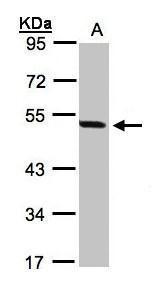
- Main image
- Experimental details
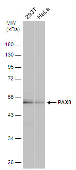
- Sample(30 ug whole cell lysate)A:293T10% SDS PAGEGTX101583 diluted at 1:1000
- Validation comment
- WB
- Submitted by
- GeneTex (provider)
- Main image
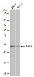
- Experimental details
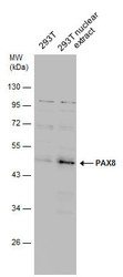
- Various whole cell extracts (30 ?g) were separated by 10% SDS-PAGE, and the membrane was blotted with PAX8 antibody (GTX101583) diluted at 1:1000. The HRP-conjugated anti-rabbit IgG antibody (GTX213110-01) was used to detect the primary antibody.
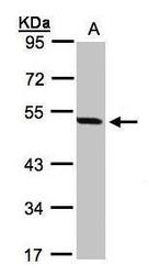
- Submitted by
- GeneTex (provider)
- Main image
- Experimental details
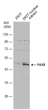
- 293T whole cell and nuclear extracts (30 ?g) were separated by 10% SDS-PAGE, and the membrane was blotted with PAX8 antibody (GTX101583) diluted at 1:1000.
Supportive validation
- Submitted by
- GeneTex (provider)
- Main image

- Experimental details
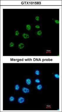
- Immunofluorescence analysis of paraformaldehyde-fixed A549, using PAX8(GTX101583) antibody at 1:200 dilution.
Supportive validation
- Submitted by
- GeneTex (provider)
- Main image
- Experimental details
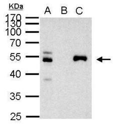
- PAX8 antibody immunoprecipitates PAX8 protein in IP experiments. IP Sample: 1000 ?g 293T whole cell lysate/extract A. 40 £gg 293T whole cell lysate/extract B. Control with 2 £gg of preimmune rabbit IgG C. Immunoprecipitation of PAX8 protein by 2 £gg of PAX8 antibody (GTX101583) 10% SDS-PAGE The immunoprecipitated PAX8 protein was detected by PAX8 antibody (GTX101583) diluted at 1:1000. EasyBlot anti-rabbit IgG (GTX221666-01) was used as a secondary reagent.

Supportive validation
- Submitted by
- GeneTex (provider)
- Main image
- Experimental details
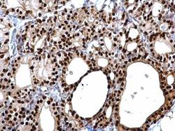
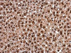
- PAX8 antibody detects PAX8 protein at cytosol and nucleus on rat thyroid gland by immunohistochemical analysis. Sample: Paraffin-embedded rat thyroid gland. PAX8 antibody (GTX101583) dilution: 1:500.
- Submitted by
- GeneTex (provider)
- Main image
- Experimental details
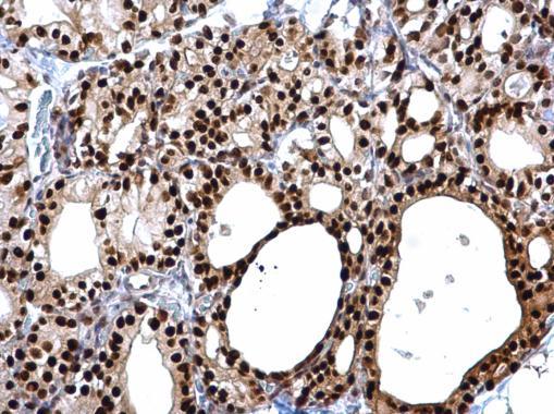
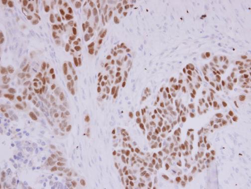
- PAX8 antibody detects PAX8 protein at nucleolus by immunohistochemical analysis. Sample: Paraffin-embedded DLD1 xenograft . PAX8 antibody (GTX101583) diluted at 1:500.
- Submitted by
- GeneTex (provider)
- Main image
- Experimental details
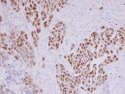
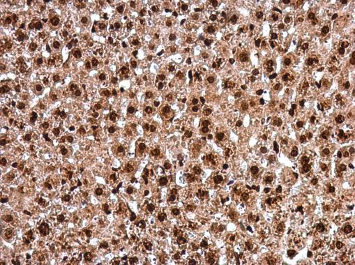
- PAX8 antibody detects PAX8 protein at nucleus in mouse liver by immunohistochemical analysis. Sample: Paraffin-embedded mouse liver. PAX8 antibody (GTX101583) diluted at 1:500.
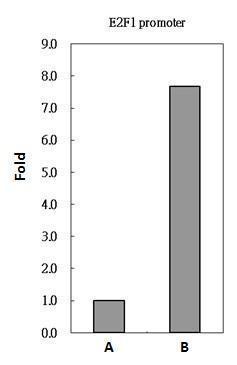
Supportive validation
- Submitted by
- GeneTex (provider)
- Main image
- Experimental details
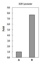
- PAX8 antibody immunoprecipitates PAX8 protein-DNA in ChIP experiments. ChIP Sample: 293T whole cell lysate/extract A. 5 £gg preimmune rabbit IgG B. 5 £gg of PAX8 antibody (GTX101583) The precipitated DNA was detected by PCR with primer set targeting to E2F1 promoter.