Explore
 Validate
 Learn
Learn Western blot
 Immunocytochemistry
Antibody data
- Antibody Data
- Antigen structure
- References [2]
- Comments [0]
- Validations
- Immunocytochemistry [1]
Submit
Validation data
Reference
Comment
Report error
- Product number
- AF3824 - Provider product page

- Provider
- R&D Systems
- Product name
- Human Integrin beta 5 Antibody
- Antibody type
- Polyclonal
- Description
- Antigen Affinity-purified. Detects human Integrin beta 5 in direct ELISAs and Western blots. In direct ELISAs, approximately 50% cross-reactivity with recombinant mouse (rm) Integrin beta 5 is observed, and less than 5% cross-reactivity with recombinant human (rh) Integrin beta 1, rhIntegrin beta 3, rhIntegrin beta 4, rhIntegrin beta 6 and rmIntegrin beta 7 is observed.
- Reactivity
- Human
- Host
- Sheep
- Conjugate
- Unconjugated
- Antigen sequence
P18084- Isotype
- IgG
- Vial size
- 100 ug
- Concentration
- LYOPH
- Storage
- Use a manual defrost freezer and avoid repeated freeze-thaw cycles. 12 months from date of receipt, -20 to -70 °C as supplied. 1 month, 2 to 8 °C under sterile conditions after reconstitution. 6 months, -20 to -70 °C under sterile conditions after reconstitution.
Submitted references Targeting cadherin-17 inactivates Ras/Raf/MEK/ERK signaling and inhibits cell proliferation in gastric cancer.
Development of a method for the purification and culture of rodent astrocytes.
Lin Z, Zhang C, Zhang M, Xu D, Fang Y, Zhou Z, Chen X, Qin N, Zhang X
PloS one 2014;9(1):e85296
PloS one 2014;9(1):e85296
Development of a method for the purification and culture of rodent astrocytes.
Foo LC, Allen NJ, Bushong EA, Ventura PB, Chung WS, Zhou L, Cahoy JD, Daneman R, Zong H, Ellisman MH, Barres BA
Neuron 2011 Sep 8;71(5):799-811
Neuron 2011 Sep 8;71(5):799-811
No comments: Submit comment
Supportive validation
- Submitted by
- R&D Systems (provider)
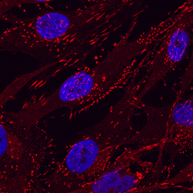
- Main image

- Experimental details
- Integrin beta 5 in MG-63 Human Cell Line. Integrin beta 5 was detected in immersion fixed MG-63 human osteosarcoma cell line using Sheep Anti-Human Integrin beta 5 Antigen Affinity-purified Polyclonal Antibody (Catalog # AF3824) at 10 µg/mL for 3 hours at room temperature. Cells were stained using the NorthernLights™ 557-conjugated Anti-Sheep IgG Secondary Antibody (red; Catalog # NL010) and counterstained with DAPI(blue). Specific staining was localized to cytoplasm. View our protocol for Fluorescent ICC Staining of Cells on Coverslips.