Explore
 Validate
 Learn
LearnPA5-19895
antibody from Invitrogen Antibodies
Targeting: TNFRSF10B
CD262, DR5, KILLER, TRAIL-R2, TRAILR2, TRICK2A, TRICKB
 Western blot
Antibody data
- Antibody Data
- Antigen structure
- References [0]
- Comments [0]
- Validations
- Western blot [7]
- Immunocytochemistry [2]
- Immunohistochemistry [7]
Submit
Validation data
Reference
Comment
Report error
- Product number
- PA5-19895 - Provider product page

- Provider
- Invitrogen Antibodies
- Product name
- TRAIL-R2 (DR5) Polyclonal Antibody
- Antibody type
- Polyclonal
- Antigen
- Synthetic peptide
- Description
- A suggested positive control is Hela cell lysate. PA5-19895 can be used with blocking peptide PEP-0021. The PA5-19895 immunogen is located within the last 50 amino acids of DR5. Predicted molecular ~ 48kD and 45kD. In Western blot applications, this antibody has been observed to detect a band at: 48kD and 45kD
- Reactivity
- Human, Mouse, Rat
- Host
- Rabbit
- Isotype
- IgG
- Vial size
- 100 µg
- Concentration
- 1 mg/mL
- Storage
- 4° C
No comments: Submit comment
Supportive validation
- Submitted by
- Invitrogen Antibodies (provider)
- Main image

- Experimental details
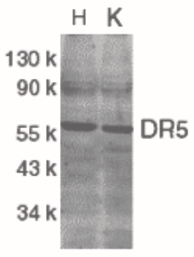
- Western blot analysis of HeLa (H) and K562 (K) cell lysates using a CD262/TRAIL Receptor 2 DR5 polyclonal antibody (Product # PA5-19895) at 2 µg/mL.
- Submitted by
- Invitrogen Antibodies (provider)
- Main image

- Experimental details
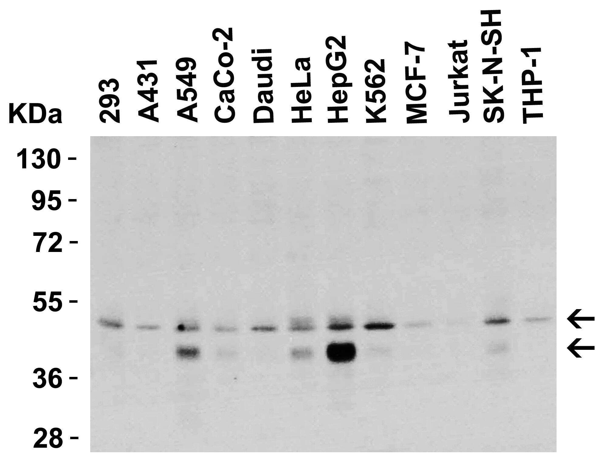
- Western Blot Validation in Human Cell Lines. Loading: 15 µg of lysates per lane. Antibodies: TRAIL-R2 (DR5) Polyclonal Antibody (Product # PA5-19895) (0.5 µg/mL), 1h incubation at RT in 0.05 NFDM/TBST. Secondary: Goat anti-rabbit IgG HRP conjugate at 1:10,000 dilution.
- Submitted by
- Invitrogen Antibodies (provider)
- Main image

- Experimental details
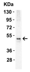
- Western Blot Validation in Human HepG2 Cells. Loading: 15 µg of lysates per lane. Antibodies: TRAIL-R2 (DR5) Polyclonal Antibody (Product # PA5-19895) 1h incubation at RT in 0.05 NFDM/TBST. Secondary: Goat anti-rabbit IgG HRP conjugate at 1:10,000 dilution. Lane 1: 1 µg/mL Lane 2: 2 µg/mL Lane 3: 4 µg/mL

- Submitted by
- Invitrogen Antibodies (provider)
- Main image

- Experimental details
- Western Blot Validation in Mouse and Rat Cell Lines. Loading: 15 µg of lysates per lane. Antibodies: TRAIL-R2 (DR5) Polyclonal Antibody (Product # PA5-19895) (2 µg/mL), 1h incubation at RT in 0.05 NFDM/TBST. Secondary: Goat anti-rabbit IgG HRP

- Submitted by
- Invitrogen Antibodies (provider)
- Main image

- Experimental details
- Western Blot Validation in Mouse Cell Lines. Loading: 15 µg of lysates per lane. Antibodies: TRAIL-R2 (DR5) Polyclonal Antibody (Product # PA5-19895) (1 µg/mL), 1h incubation at RT in 0.05 NFDM/TBST. Secondary: Goat anti-rabbit IgG HRP conjugate at 1:10,000 dilution.

- Submitted by
- Invitrogen Antibodies (provider)
- Main image

- Experimental details
- Western Blot Validation in Mouse Heart. Loading: 15 µg of lysatesper lane. Antibodies: TRAIL-R2 (DR5) Polyclonal Antibody (Product # PA5-19895) (1 µg/mL), 1h incubation at RT in 0.05 NFDM/TBST. Secondary: Goat anti-rabbit IgG HRP conjugate at 1:10,000 dilution.

- Submitted by
- Invitrogen Antibodies (provider)
- Main image
- Experimental details
- Western Blot Validation in Rat Skeletal Muscle. Loading: 15 µg of lysate per lane. Antibodies: TRAIL-R2 (DR5) Polyclonal Antibody (Product # PA5-19895) (1 µg/mL), 1h incubation at RT in 0.05 NFDM/TBST. Secondary: Goat anti-rabbit IgG HRP conjugate at 1:10,000 dilution.

Supportive validation
- Submitted by
- Invitrogen Antibodies (provider)
- Main image

- Experimental details
- Immunofluorescent analysis of HeLa cells using a CD262/TRAIL Receptor 2 DR5 polyclonal antibody (Product # PA5-19895) at a 20 µg/mL dilution.

- Submitted by
- Invitrogen Antibodies (provider)
- Main image

- Experimental details
- Immunofluorescent analysis of 4% paraformaldehyde-fixed human HepG2 cells labeling DR5 with TRAIL-R2 (DR5) Polyclonal Antibody (Product # PA5-19895) at 5 µg/mL, followed by goat anti-rabbit IgG secondary antibody at 1:500 dilution (green) and DAPI (blue).

Supportive validation
- Submitted by
- Invitrogen Antibodies (provider)
- Main image

- Experimental details
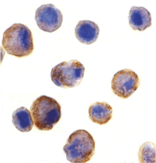
- Immunocytochemistry staining of HeLa cells using a CD262/TRAIL Receptor 2 DR5 polyclonal antibody (Product # PA5-19895) at a 5 µg/mL dilution.
- Submitted by
- Invitrogen Antibodies (provider)
- Main image

- Experimental details
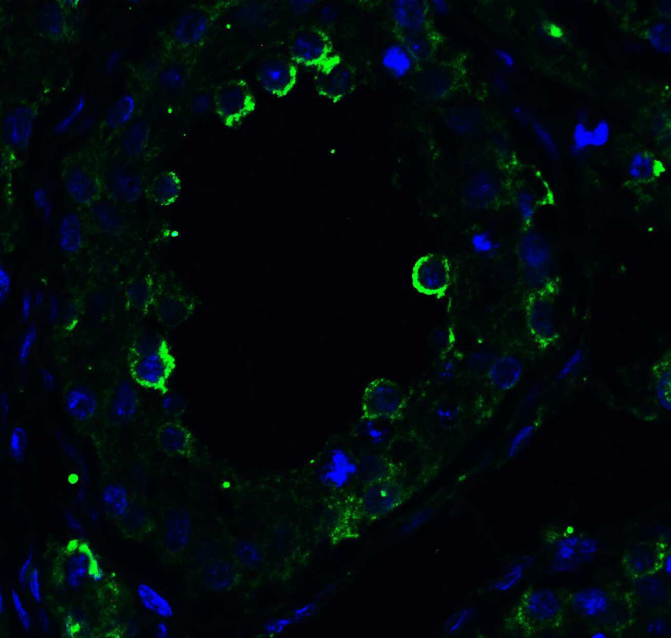
- Immunofluorescent analysis of 4% paraformaldehyde-fixed human testis tissue labeling DR5 with TRAIL-R2 (DR5) Polyclonal Antibody (Product # PA5-19895) at 10 µg/mL, followed by goat anti-rabbit IgG secondary antibody at 1:500 dilution (green) and DAPI (blue).
- Submitted by
- Invitrogen Antibodies (provider)
- Main image

- Experimental details
- Immunofluorescent analysis of 4% paraformaldehyde-fixed rat brain tissue labeling DR5 with TRAIL-R2 (DR5) Polyclonal Antibody (Product # PA5-19895) at 5 µg/mL, followed by goat anti-rabbit IgG secondary antibody at 1:500 dilution (green) and DAPI (blue).

- Submitted by
- Invitrogen Antibodies (provider)
- Main image

- Experimental details
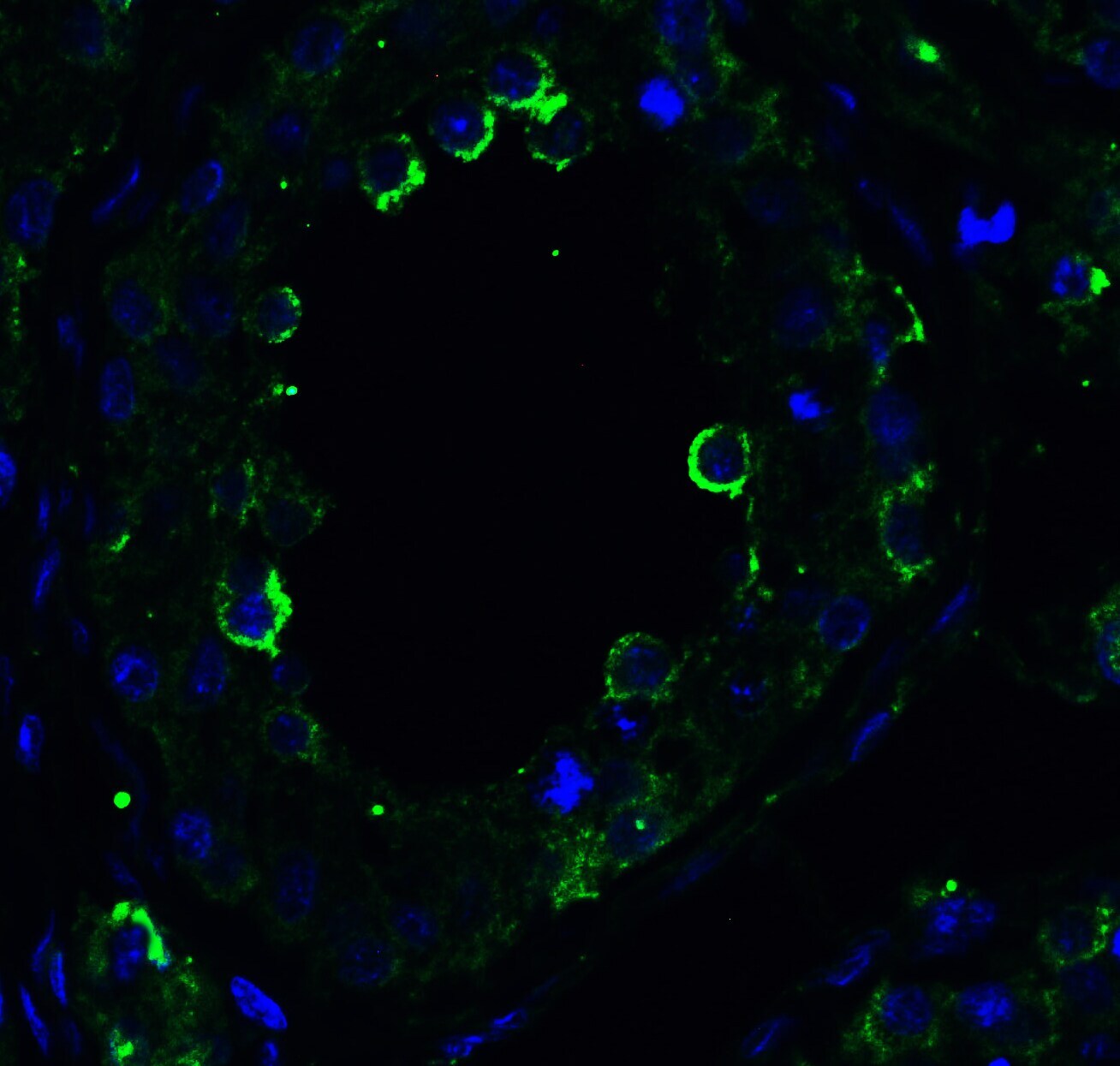
- Immunofluorescent analysis of 4% paraformaldehyde-fixed human testis tissue labeling DR5 with TRAIL-R2 (DR5) Polyclonal Antibody (Product # PA5-19895) at 10 µg/mL, followed by goat anti-rabbit IgG secondary antibody at 1:500 dilution (green) and DAPI (blue).
- Submitted by
- Invitrogen Antibodies (provider)
- Main image

- Experimental details
- Immunofluorescent analysis of 4% paraformaldehyde-fixed mouse pancreas tissue labeling DR5 with TRAIL-R2 (DR5) Polyclonal Antibody (Product # PA5-19895) at 10 µg/mL, followed by goat anti-rabbit IgG secondary antibody at 1:500 dilution (green) and DAPI (blue).

- Submitted by
- Invitrogen Antibodies (provider)
- Main image

- Experimental details
- Immunofluorescent analysis of 4% paraformaldehyde-fixed rat brain tissue labeling DR5 with TRAIL-R2 (DR5) Polyclonal Antibody (Product # PA5-19895) at 5 µg/mL, followed by goat anti-rabbit IgG secondary antibody at 1:500 dilution (green) and DAPI (blue).

- Submitted by
- Invitrogen Antibodies (provider)
- Main image

- Experimental details
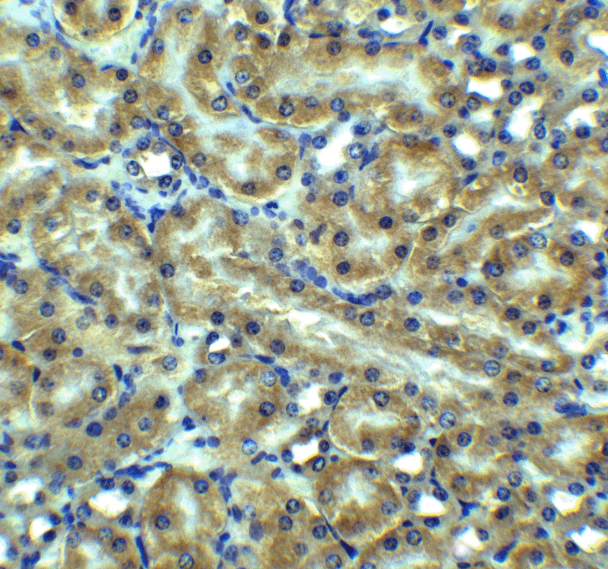
- Immunohistochemical analysis of paraffin-embedded mouse kidney tissue using TRAIL-R2 (DR5) Polyclonal Antibody (Product # PA5-19895) at 5 µg/mL. Tissue was fixed with formaldehyde and blocked with 0.1 serum for 1 h at RT; antigen retrieval was by heat mediation with a citrate buffer (pH6). Samples were incubated with primary antibody overnight at 4˚C. A goat anti-rabbit IgG H&L (HRP) at 1/250 was used as secondary. Counter stained with Hematoxylin.